


3GD8
Crystal Structure of Human Aquaporin 4 at 1.8 and its Mechanism of Conductance
- PDB DOI: https://doi.org/10.2210/pdb3GD8/pdb
- Classification: MEMBRANE PROTEIN
- Organism(s): Homo sapiens
- Expression System: Komagataella pastoris
- Mutation(s): No
- Membrane Protein: Yes OPMPDBTMMemProtMDmpstruc
- Deposited: 2009-02-23 Released: 2009-03-31
Experimental Data Snapshot
- Method: X-RAY DIFFRACTION
- Resolution: 1.80 Å
- R-Value Free: 0.165
- R-Value Work: 0.160
- R-Value Observed: 0.160

This is version 1.3 of the entry. See complete history.

Macromolecules
Find similar proteins by:
(by identity cutoff) | 3D Structure
Entity ID: 1 | |||||
|---|---|---|---|---|---|
| Molecule | Chains | Sequence Length | Organism | Details | Image |
| Aquaporin-4 | 223 | Homo sapiens | Mutation(s): 0 Gene Names: AQP4 Membrane Entity: Yes | 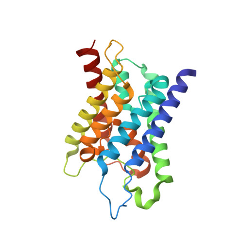 | |
UniProt & NIH Common Fund Data Resources | |||||
Find proteins for P55087 (Homo sapiens) Explore P55087 Go to UniProtKB: P55087 | |||||
PHAROS: P55087 GTEx: ENSG00000171885 | |||||
Entity Groups | |||||
| Sequence Clusters | 30% Identity50% Identity70% Identity90% Identity95% Identity100% Identity | ||||
| UniProt Group | P55087 | ||||
Sequence AnnotationsExpand | |||||
| |||||
Small Molecules
| Ligands 2 Unique | |||||
|---|---|---|---|---|---|
| ID | Chains | Name / Formula / InChI Key | 2D Diagram | 3D Interactions | |
| BOG Query on BOG | G [auth A] | octyl beta-D-glucopyranoside C14 H28 O6 HEGSGKPQLMEBJL-RKQHYHRCSA-N |  | ||
| GOL Query on GOL | B [auth A], C [auth A], D [auth A], E [auth A], F [auth A] | GLYCEROL C3 H8 O3 PEDCQBHIVMGVHV-UHFFFAOYSA-N |  | ||
Experimental Data & Validation
Experimental Data
- Method: X-RAY DIFFRACTION
- Resolution: 1.80 Å
- R-Value Free: 0.165
- R-Value Work: 0.160
- R-Value Observed: 0.160
- Space Group: P 4 21 2
Unit Cell:
| Length ( Å ) | Angle ( ˚ ) |
|---|---|
| a = 82.058 | α = 90 |
| b = 82.058 | β = 90 |
| c = 76.353 | γ = 90 |
| Software Name | Purpose |
|---|---|
| Blu-Ice | data collection |
| PHASER | phasing |
| REFMAC | refinement |
| HKL-2000 | data reduction |
| HKL-2000 | data scaling |
Entry History
Deposition Data
- Released Date: 2009-03-31 Deposition Author(s): Ho, J.D., Yeh, R., Sandstrom, A., Chorny, I., Harries, W.E.C., Robbins, R.A., Miercke, L.J.W., Stroud, R.M., Center for Structures of Membrane Proteins (CSMP)
Revision History (Full details and data files)
- Version 1.0: 2009-03-31
Type: Initial release - Version 1.1: 2011-07-13
Changes: Advisory, Version format compliance - Version 1.2: 2020-07-29
Type: Remediation
Reason: Carbohydrate remediation
Changes: Data collection, Derived calculations, Structure summary - Version 1.3: 2024-02-21
Changes: Data collection, Database references, Structure summary
