Electrostatic interactions contribute to the folded-back conformation of wild type human factor H.
Okemefuna, A.I., Nan, R., Gor, J., Perkins, S.J.(2009) J Mol Biol 391: 98-118
- PubMed: 19505476
- DOI: https://doi.org/10.1016/j.jmb.2009.06.010
- Primary Citation of Related Structures:
3GAU, 3GAV, 3GAW - PubMed Abstract:
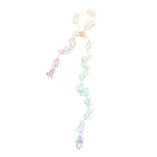
Factor H (FH), a major serum regulator of C3b in the complement alternative pathway, is composed of 20 short complement regulator (SCR) domains. Earlier solution structures for FH showed that this has a folded-back domain arrangement and exists as oligomers. To clarify the molecular basis for this, analytical ultracentrifugation and X-ray scattering studies of native FH were performed as a function of NaCl concentration and pH. The sedimentation coefficient for the FH monomer decreased from 5.7 S to 5.3 S with increase in NaCl concentration, showing that weak electrostatic inter-domain interactions affect its folded-back structure. FH became more elongated at pH 9.4, showing the involvement of histidine residue(s) in its folded-back structure. Similar studies of partially deglycosylated FH suggested that oligosaccharides were not significant in determining the FH domain structure. The formation of FH oligomers decreased with increased NaCl concentration, indicating that electrostatic interactions also affect this. X-ray scattering showed that the maximum length of FH increased from 32 nm in low salt to 38 nm in high salt. Constrained X-ray scattering modelling was used to generate significantly improved FH molecular structures at medium resolution. In 50 mM NaCl, the modelled structures showed that inter-SCR domain contacts are likely, while these contacts are fewer in 250 mM NaCl. The results of this study show that the conformation of FH is affected by its local environment, and this may be important for its interactions with C3b and when bound to polyanionic cell surfaces.
Organizational Affiliation:
Institute of Structural and Molecular Biology, Division of Biosciences, Darwin Building, University College London, Gower Street, London, UK.