3G6Y

Ternary complex of DNA Polymerase iota:DNA:dTTP with an abasic site at the templating position


Experimental Data Snapshot

  • Method: X-RAY DIFFRACTION
  • Resolution: 2.10 Å
  • R-Value Free: 0.253 
  • R-Value Work: 0.220 
  • R-Value Observed: 0.223 

wwPDB Validation   3D Report Full Report


Ligand Structure Quality Assessment 


This is version 1.4 of the entry. See complete history


Literature

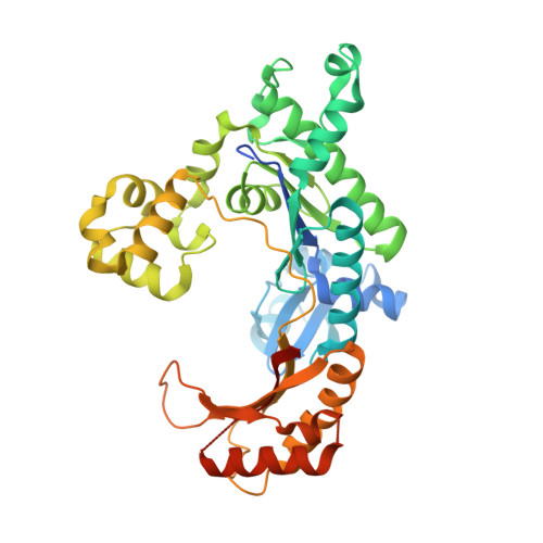
DNA Synthesis across an Abasic Lesion by Human DNA Polymerase iota

Nair, D.T.Johnson, R.E.Prakash, L.Prakash, S.Aggarwal, A.K.

(2009) Structure 17: 530-537

  • DOI: https://doi.org/10.1016/j.str.2009.02.015
  • Primary Citation of Related Structures:  
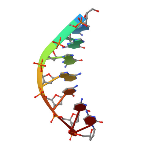
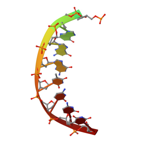
    3G6V, 3G6X, 3G6Y

  • PubMed Abstract: 

    Abasic sites are among the most abundant DNA lesions formed in human cells, and they present a strong block to replication. DNA polymerase iota (Poliota) is one of the few DNA Pols that does not follow the A-rule opposite an abasic site. We present here three structures of human Poliota in complex with DNAs containing an abasic lesion and dGTP, dTTP, or dATP as the incoming nucleotide. The structures reveal a mechanism of translesion synthesis across an abasic lesion that differs from that in other Pols. Both the abasic lesion and the incoming dNTPs are intrahelical and are closely apposed across a constricted active site cleft. The dNTPs partake in distinct networks of hydrogen bonds in the "void" opposite the lesion. These different patterns of hydrogen bonds, as well as stacking interactions, may underlie Poliota's small preference for insertion of dGTP over other nucleotides opposite this common lesion.


  • Organizational Affiliation

    Department of Structural and Chemical Biology, Mount Sinai School of Medicine, New York, NY 10029, USA.


Macromolecules

Find similar proteins by:  (by identity cutoff)  |  3D Structure
Entity ID: 1
MoleculeChains Sequence LengthOrganismDetailsImage
DNA polymerase iota420Homo sapiensMutation(s): 0 
Gene Names: POLIRAD30B
EC: 2.7.7.7
UniProt & NIH Common Fund Data Resources
Find proteins for Q9UNA4 (Homo sapiens)
Explore Q9UNA4 
Go to UniProtKB:  Q9UNA4
PHAROS:  Q9UNA4
GTEx:  ENSG00000101751 
Entity Groups  
Sequence Clusters30% Identity50% Identity70% Identity90% Identity95% Identity100% Identity
UniProt GroupQ9UNA4
Sequence Annotations
Expand
  • Reference Sequence

Find similar nucleic acids by:  Sequence   |   3D Structure  

Entity ID: 2
MoleculeChains LengthOrganismImage
Primer DNA strandB [auth P]7N/A
Sequence Annotations
Expand
  • Reference Sequence

Find similar nucleic acids by:  Sequence   |   3D Structure  

Entity ID: 3
MoleculeChains LengthOrganismImage
Template DNA strandC [auth T]11N/A
Sequence Annotations
Expand
  • Reference Sequence
Experimental Data & Validation

Experimental Data

  • Method: X-RAY DIFFRACTION
  • Resolution: 2.10 Å
  • R-Value Free: 0.253 
  • R-Value Work: 0.220 
  • R-Value Observed: 0.223 
  • Space Group: P 65 2 2
Unit Cell:
Length ( Å )Angle ( ˚ )
a = 98.114α = 90
b = 98.114β = 90
c = 202.823γ = 120
Software Package:
Software NamePurpose
REFMACrefinement
AMoREphasing
CNSrefinement
JDirectordata collection
HKL-2000data reduction
HKL-2000data scaling

Structure Validation

View Full Validation Report


Ligand Structure Quality Assessment 


Entry History 

Deposition Data

Revision History  (Full details and data files)

  • Version 1.0: 2009-05-12
    Type: Initial release
  • Version 1.1: 2011-07-13
    Changes: Advisory, Refinement description, Version format compliance
  • Version 1.2: 2017-11-01
    Changes: Refinement description
  • Version 1.3: 2019-07-24
    Changes: Data collection, Derived calculations, Refinement description
  • Version 1.4: 2023-09-06
    Changes: Data collection, Database references, Derived calculations, Refinement description