


Queen bee pheromone binding protein pH induced domain-swapping favors pheromone release
Pesenti, M.E., Spinelli, S., Bezirard, V., Briand, L., Pernollet, J.C., Tegoni, M., Cambillau, C.To be published.
Experimental Data Snapshot
Entity ID: 1 | |||||
|---|---|---|---|---|---|
| Molecule | Chains | Sequence Length | Organism | Details | Image |
| Pheromone-binding protein ASP1 | 119 | Apis mellifera | Mutation(s): 0 | 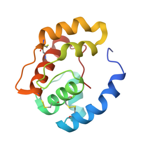 | |
UniProt | |||||
Find proteins for Q9U9J6 (Apis mellifera) Explore Q9U9J6 Go to UniProtKB: Q9U9J6 | |||||
Entity Groups | |||||
| Sequence Clusters | 30% Identity50% Identity70% Identity90% Identity95% Identity100% Identity | ||||
| UniProt Group | Q9U9J6 | ||||
Sequence AnnotationsExpand | |||||
| |||||
| Ligands 3 Unique | |||||
|---|---|---|---|---|---|
| ID | Chains | Name / Formula / InChI Key | 2D Diagram | 3D Interactions | |
| CMJ Query on CMJ | B [auth A] | (20S)-20-methyldotetracontane C43 H88 RQRQHJNVHOWJHC-QLKFWGTOSA-N |  | ||
| GOL Query on GOL | C [auth A] | GLYCEROL C3 H8 O3 PEDCQBHIVMGVHV-UHFFFAOYSA-N |  | ||
| CL Query on CL | D [auth A] | CHLORIDE ION Cl VEXZGXHMUGYJMC-UHFFFAOYSA-M |  | ||
| Length ( Å ) | Angle ( ˚ ) |
|---|---|
| a = 82.879 | α = 90 |
| b = 84.21 | β = 90 |
| c = 46.831 | γ = 90 |
| Software Name | Purpose |
|---|---|
| ADSC | data collection |
| REFMAC | refinement |
| MOSFLM | data reduction |
| SCALA | data scaling |
| REFMAC | phasing |
RCSB PDB (citation) is hosted by
RCSB PDB is a member of the

