3FCX

Crystal structure of human esterase D


Experimental Data Snapshot

  • Method: X-RAY DIFFRACTION
  • Resolution: 1.50 Å
  • R-Value Free: 0.196 
  • R-Value Work: 0.176 
  • R-Value Observed: 0.177 

wwPDB Validation   3D Report Full Report


This is version 1.3 of the entry. See complete history


Literature

Crystal structure of human esterase D: a potential genetic marker of retinoblastoma

Wu, D.Li, Y.Song, G.Zhang, D.Shaw, N.Liu, Z.J.

(2009) FASEB J 23: 1441-1446

  • DOI: https://doi.org/10.1096/fj.08-125286
  • Primary Citation of Related Structures:  
    3FCX

  • PubMed Abstract: 

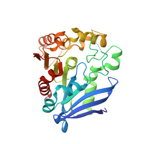
    Retinoblastoma (RB), a carcinoma of the retina, is caused by mutations in the long arm of chromosome 13, band 13q14. The esterase D (ESD) gene maps at a similar location as the RB gene locus and therefore serves as a potential marker for the prognosis of retinoblastoma. Because very little is known about the structure and function of ESD, we determined the 3-dimensional structure of the enzyme at 1.5 A resolution using X-ray crystallography. ESD shows a single domain with an alpha/beta-hydrolase fold. A number of insertions are observed in the canonical alpha/beta-hydrolase fold. The active site is located in a positively charged, shallow cleft on the surface lined by a number of aromatic residues. Superimposition studies helped identify the typical catalytic triad residues--Ser-153, His264, and Asp230--involved in catalysis. Mutagenesis of any of the catalytic triad residues to alanine abolished the enzyme activity. Backbone amides of Leu54 and Met150 are involved in the formation of the oxyanion hole. Interestingly, a M150A mutation increased the enzyme activity by 62%. The structure of human ESD determined in this study will aid the elucidation of the physiological role of the enzyme in the human body and will assist in the early diagnosis of retinoblastoma.


  • Organizational Affiliation

    National Laboratory of Biomacromolecules, Institute of Biophysics, 15 Datun Lu, Beijing 100101, China.


Macromolecules
Find similar proteins by:  (by identity cutoff)  |  3D Structure
Entity ID: 1
MoleculeChains Sequence LengthOrganismDetailsImage
S-formylglutathione hydrolase
A, B
282Homo sapiensMutation(s): 1 
Gene Names: ESD
EC: 3.1.2.12
UniProt & NIH Common Fund Data Resources
Find proteins for P10768 (Homo sapiens)
Explore P10768 
Go to UniProtKB:  P10768
PHAROS:  P10768
GTEx:  ENSG00000139684 
Entity Groups  
Sequence Clusters30% Identity50% Identity70% Identity90% Identity95% Identity100% Identity
UniProt GroupP10768
Sequence Annotations
Expand
  • Reference Sequence
Experimental Data & Validation

Experimental Data

  • Method: X-RAY DIFFRACTION
  • Resolution: 1.50 Å
  • R-Value Free: 0.196 
  • R-Value Work: 0.176 
  • R-Value Observed: 0.177 
  • Space Group: P 1 21 1
Unit Cell:
Length ( Å )Angle ( ˚ )
a = 51.542α = 90
b = 70.724β = 108.84
c = 65.014γ = 90
Software Package:
Software NamePurpose
HKL-2000data collection
PHASERphasing
REFMACrefinement
HKL-2000data reduction
HKL-2000data scaling

Structure Validation

View Full Validation Report


Entry History 

Deposition Data

Revision History  (Full details and data files)

  • Version 1.0: 2008-12-09
    Type: Initial release
  • Version 1.1: 2011-07-13
    Changes: Version format compliance
  • Version 1.2: 2021-11-10
    Changes: Database references, Derived calculations
  • Version 1.3: 2023-11-01
    Changes: Data collection, Refinement description