3F7B

Crystal Structure of soluble domain of CA4 in complex with small molecule.


Experimental Data Snapshot

  • Method: X-RAY DIFFRACTION
  • Resolution: 2.05 Å
  • R-Value Free: 0.252 
  • R-Value Work: 0.191 
  • R-Value Observed: 0.194 

wwPDB Validation   3D Report Full Report


Ligand Structure Quality Assessment 


This is version 1.2 of the entry. See complete history


Literature

Thioether benzenesulfonamide inhibitors of carbonic anhydrases II and IV: structure-based drug design, synthesis, and biological evaluation.

Vernier, W.Chong, W.Rewolinski, D.Greasley, S.Pauly, T.Shaw, M.Dinh, D.Ferre, R.A.Nukui, S.Ornelas, M.Reyner, E.

(2010) Bioorg Med Chem 18: 3307-3319

  • DOI: https://doi.org/10.1016/j.bmc.2010.03.014
  • Primary Citation of Related Structures:  
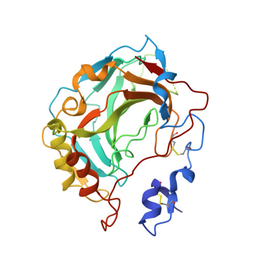
    3F7B, 3F7U

  • PubMed Abstract: 

    A novel series of potent thioether benzenesulfonamide inhibitors of carbonic anhydrases II and IV was discovered using structure-based drug design. Synthesis, structure-activity relationship, and optimization of physicochemical properties are described. Low nanomolar potency was achieved, and selected compounds with improved thermodynamic solubility showed promising in vitro inhibition of carbonic anhydrase activity in rabbit iris ciliary body homogenate.


  • Organizational Affiliation

    Pfizer Global Research and Development, La Jolla Laboratories, 10770 Science Center Drive, San Diego, CA 92121, USA. william.f.vernier@pfizer.com


Macromolecules
Find similar proteins by:  (by identity cutoff)  |  3D Structure
Entity ID: 1
MoleculeChains Sequence LengthOrganismDetailsImage
Carbonic anhydrase 4
A, B
266Homo sapiensMutation(s): 0 
Gene Names: CA4
EC: 4.2.1.1
UniProt & NIH Common Fund Data Resources
Find proteins for P22748 (Homo sapiens)
Explore P22748 
Go to UniProtKB:  P22748
PHAROS:  P22748
GTEx:  ENSG00000167434 
Entity Groups  
Sequence Clusters30% Identity50% Identity70% Identity90% Identity95% Identity100% Identity
UniProt GroupP22748
Sequence Annotations
Expand
  • Reference Sequence
Small Molecules
Binding Affinity Annotations 
IDSourceBinding Affinity
AG5 Binding MOAD:  3F7B IC50: 64 (nM) from 1 assay(s)
BindingDB:  3F7B IC50: 64 (nM) from 1 assay(s)
PDBBind:  3F7B IC50: 64 (nM) from 1 assay(s)
Experimental Data & Validation

Experimental Data

  • Method: X-RAY DIFFRACTION
  • Resolution: 2.05 Å
  • R-Value Free: 0.252 
  • R-Value Work: 0.191 
  • R-Value Observed: 0.194 
  • Space Group: C 1 2 1
Unit Cell:
Length ( Å )Angle ( ˚ )
a = 125.91α = 90
b = 128.556β = 99.88
c = 46.365γ = 90
Software Package:
Software NamePurpose
HKL-2000data collection
AMoREphasing
REFMACrefinement
HKL-2000data reduction
HKL-2000data scaling

Structure Validation

View Full Validation Report


Ligand Structure Quality Assessment 


Entry History 

Deposition Data

Revision History  (Full details and data files)

  • Version 1.0: 2009-09-22
    Type: Initial release
  • Version 1.1: 2011-07-13
    Changes: Version format compliance
  • Version 1.2: 2023-09-06
    Changes: Data collection, Database references, Derived calculations, Refinement description