3ESK

Structure of HOP TPR2A domain in complex with the non-cognate Hsc70 peptide ligand


Experimental Data Snapshot

  • Method: X-RAY DIFFRACTION
  • Resolution: 2.05 Å
  • R-Value Free: 0.244 
  • R-Value Work: 0.177 
  • R-Value Observed: 0.183 

wwPDB Validation   3D Report Full Report


This is version 1.3 of the entry. See complete history


Literature

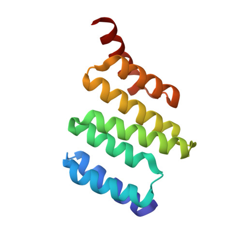
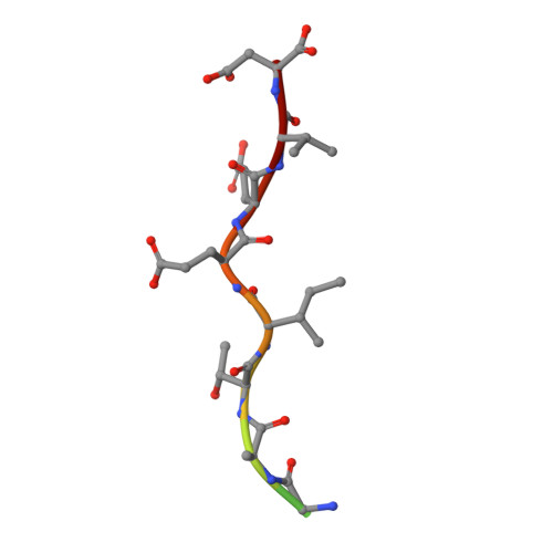
Electrostatic interactions of Hsp-organizing protein tetratricopeptide domains with Hsp70 and Hsp90: computational analysis and protein engineering.

Kajander, T.Sachs, J.N.Goldman, A.Regan, L.

(2009) J Biol Chem 284: 25364-25374

  • DOI: https://doi.org/10.1074/jbc.M109.033894
  • Primary Citation of Related Structures:  
    3ESK

  • PubMed Abstract: 

    The Hsp-organizing protein (HOP) binds to the C termini of the chaperones Hsp70 and Hsp90, thus bringing them together so that substrate proteins can be passed from Hsp70 to Hsp90. Because Hsp90 is essential for the correct folding and maturation of many oncogenic proteins, it has become a significant target for anti-cancer drug design. HOP binds to Hsp70 and Hsp90 via two independent tetratricopeptide (TPR) domains, TPR1 and TPR2A, respectively. We have analyzed ligand binding using Poisson-Boltzmann continuum electrostatic calculations, free energy perturbation, molecular dynamics simulations, and site-directed mutagenesis to delineate the contribution of different interactions to the affinity and specificity of the TPR-peptide interactions. We found that continuum electrostatic calculations could be used to guide protein design by removing unfavorable interactions to increase binding affinity, with an 80-fold increase in affinity for TPR2A. Contributions at buried charged residues, however, were better predicted by free energy perturbation calculations. We suggest using a combination of the two approaches for increasing the accuracy of results, with free energy perturbation calculations used only at selected buried residues of the ligand binding pocket. Finally we present the crystal structure of TPR2A in complex with its non-cognate Hsp70 ligand, which provides insight on the origins of specificity in TPR domain-peptide recognition.


  • Organizational Affiliation

    Department of Molecular Biophysics and Biochemistry, Yale University, New Haven, Connecticut 06511, USA. tommi.kajander@helsinki.fi


Macromolecules
Find similar proteins by:  (by identity cutoff)  |  3D Structure
Entity ID: 1
MoleculeChains Sequence LengthOrganismDetailsImage
Stress-induced-phosphoprotein 1129Homo sapiensMutation(s): 0 
Gene Names: STIP1
UniProt & NIH Common Fund Data Resources
Find proteins for P31948 (Homo sapiens)
Explore P31948 
Go to UniProtKB:  P31948
PHAROS:  P31948
GTEx:  ENSG00000168439 
Entity Groups  
Sequence Clusters30% Identity50% Identity70% Identity90% Identity95% Identity100% Identity
UniProt GroupP31948
Sequence Annotations
Expand
  • Reference Sequence

Find similar proteins by:  Sequence   |   3D Structure  

Entity ID: 2
MoleculeChains Sequence LengthOrganismDetailsImage
Heat shock cognate 71 kDa protein12Homo sapiensMutation(s): 0 
UniProt & NIH Common Fund Data Resources
Find proteins for P11142 (Homo sapiens)
Explore P11142 
Go to UniProtKB:  P11142
PHAROS:  P11142
GTEx:  ENSG00000109971 
Entity Groups  
Sequence Clusters30% Identity50% Identity70% Identity90% Identity95% Identity100% Identity
UniProt GroupP11142
Sequence Annotations
Expand
  • Reference Sequence
Small Molecules
Ligands 1 Unique
IDChains Name / Formula / InChI Key2D Diagram3D Interactions
NI
Query on NI

Download Ideal Coordinates CCD File 
C [auth A]NICKEL (II) ION
Ni
VEQPNABPJHWNSG-UHFFFAOYSA-N
Experimental Data & Validation

Experimental Data

  • Method: X-RAY DIFFRACTION
  • Resolution: 2.05 Å
  • R-Value Free: 0.244 
  • R-Value Work: 0.177 
  • R-Value Observed: 0.183 
  • Space Group: C 1 2 1
Unit Cell:
Length ( Å )Angle ( ˚ )
a = 73.459α = 90
b = 48.023β = 91.56
c = 37.896γ = 90
Software Package:
Software NamePurpose
HKL-2000data collection
AMoREphasing
REFMACrefinement
HKL-2000data reduction
HKL-2000data scaling

Structure Validation

View Full Validation Report


Entry History 

Deposition Data

Revision History  (Full details and data files)

  • Version 1.0: 2009-07-07
    Type: Initial release
  • Version 1.1: 2011-07-13
    Changes: Version format compliance
  • Version 1.2: 2014-04-02
    Changes: Source and taxonomy
  • Version 1.3: 2023-09-06
    Changes: Data collection, Database references, Derived calculations, Refinement description