


2.2 A crystal structure of inorganic pyrophosphatase from rickettsia prowazekii (p21 form)
Seattle Structural Genomics Center for Infectious Disease (SSGCID)To be published.
Experimental Data Snapshot
wwPDB Validation 3D Report Full Report

Entity ID: 1 | |||||
|---|---|---|---|---|---|
| Molecule | Chains | Sequence Length | Organism | Details | Image |
| Inorganic pyrophosphatase | 173 | Rickettsia prowazekii | Mutation(s): 0 Gene Names: ppa, RP589 EC: 3.6.1.1 | 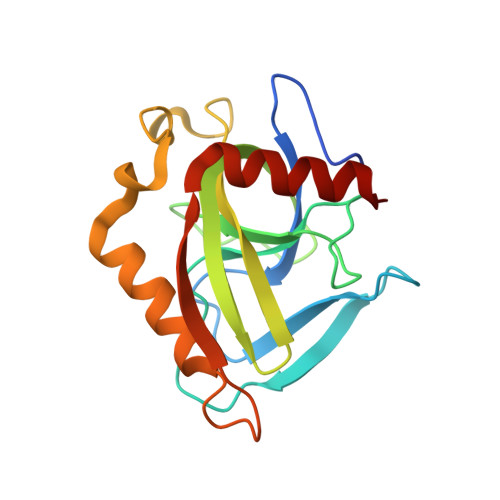 | |
UniProt | |||||
Find proteins for Q9ZCW5 (Rickettsia prowazekii (strain Madrid E)) Explore Q9ZCW5 Go to UniProtKB: Q9ZCW5 | |||||
Entity Groups | |||||
| Sequence Clusters | 30% Identity50% Identity70% Identity90% Identity95% Identity100% Identity | ||||
| UniProt Group | Q9ZCW5 | ||||
Sequence AnnotationsExpand | |||||
| |||||
| Ligands 1 Unique | |||||
|---|---|---|---|---|---|
| ID | Chains | Name / Formula / InChI Key | 2D Diagram | 3D Interactions | |
| PG4 Query on PG4 | M [auth E], N [auth L] | TETRAETHYLENE GLYCOL C8 H18 O5 UWHCKJMYHZGTIT-UHFFFAOYSA-N |  | ||
| Length ( Å ) | Angle ( ˚ ) |
|---|---|
| a = 80.96 | α = 90 |
| b = 107.35 | β = 96.05 |
| c = 128.42 | γ = 90 |
| Software Name | Purpose |
|---|---|
| SCALA | data scaling |
| MOLREP | phasing |
| REFMAC | refinement |
| PDB_EXTRACT | data extraction |
RCSB PDB (citation) is hosted by
RCSB PDB is a member of the

