3EIQ

Crystal structure of Pdcd4-eIF4A


Experimental Data Snapshot

  • Method: X-RAY DIFFRACTION
  • Resolution: 3.50 Å
  • R-Value Free: 0.269 
  • R-Value Work: 0.220 
  • R-Value Observed: 0.222 

wwPDB Validation   3D Report Full Report


This is version 1.3 of the entry. See complete history


Literature

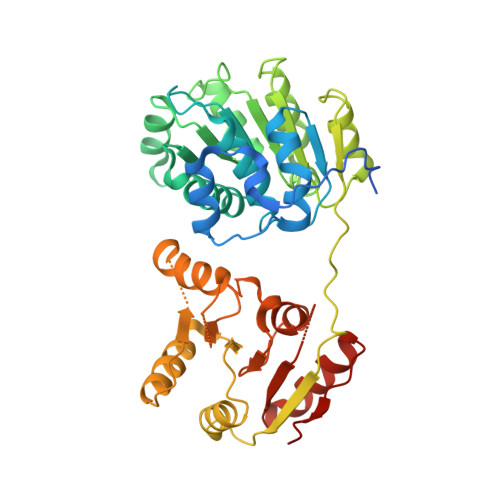
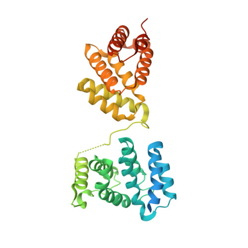
Structural basis for translational inhibition by the tumour suppressor Pdcd4

Loh, P.G.Yang, H.S.Walsh, M.A.Wang, Q.Wang, X.Cheng, Z.Liu, D.Song, H.

(2009) EMBO J 28: 274-285

  • DOI: https://doi.org/10.1038/emboj.2008.278
  • Primary Citation of Related Structures:  
    3EIJ, 3EIQ

  • PubMed Abstract: 

    Pdcd4 is a tumour suppressor protein. It inhibits translation through interaction with translation initiator eIF4A, resulting in the suppression of neoplastic transformation and tumour invasion. Here, we present the crystal structures of an N-terminal-truncated Pdcd4 in free form and in complex with eIF4A. Upon binding to eIF4A, Pdcd4 undergoes a marked conformational change to form a heterotrimeric complex with eIF4A, with one Pdcd4 binding to two eIF4A molecules in two different modes. The binding of Pdcd4 to eIF4A is required to inhibit the enzymatic activity of eIF4A, translation initiation, and AP-1-dependent transcription. Both MA3 domains are required to efficiently compete with the C-terminal domain of eIF4G (eIF4Gc) for binding to eIF4A whereas a single MA3 is sufficient to inhibit translation. Our structural and mutational analyses reveal that Pdcd4 inhibits translation initiation by trapping eIF4A in an inactive conformation, and blocking its incorporation into the eIF4F complex.


  • Organizational Affiliation

    Institute of Molecular and Cell Biology, Proteos, Singapore.


Macromolecules
Find similar proteins by:  (by identity cutoff)  |  3D Structure
Entity ID: 1
MoleculeChains Sequence LengthOrganismDetailsImage
Eukaryotic initiation factor 4A-IA,
C [auth D]
414Homo sapiensMutation(s): 0 
EC: 3.6.1
UniProt & NIH Common Fund Data Resources
Find proteins for P60842 (Homo sapiens)
Explore P60842 
Go to UniProtKB:  P60842
PHAROS:  P60842
GTEx:  ENSG00000161960 
Entity Groups  
Sequence Clusters30% Identity50% Identity70% Identity90% Identity95% Identity100% Identity
UniProt GroupP60842
Sequence Annotations
Expand
  • Reference Sequence
Find similar proteins by:  (by identity cutoff)  |  3D Structure
Entity ID: 2
MoleculeChains Sequence LengthOrganismDetailsImage
Programmed cell death protein 4B [auth C]358Mus musculusMutation(s): 0 
UniProt
Find proteins for Q61823 (Mus musculus)
Explore Q61823 
Go to UniProtKB:  Q61823
Entity Groups  
Sequence Clusters30% Identity50% Identity70% Identity90% Identity95% Identity100% Identity
UniProt GroupQ61823
Sequence Annotations
Expand
  • Reference Sequence
Experimental Data & Validation

Experimental Data

  • Method: X-RAY DIFFRACTION
  • Resolution: 3.50 Å
  • R-Value Free: 0.269 
  • R-Value Work: 0.220 
  • R-Value Observed: 0.222 
  • Space Group: P 21 3
Unit Cell:
Length ( Å )Angle ( ˚ )
a = 198.362α = 90
b = 198.362β = 90
c = 198.362γ = 90
Software Package:
Software NamePurpose
REFMACrefinement
MOSFLMdata reduction
SCALAdata scaling

Structure Validation

View Full Validation Report


Entry History 

Deposition Data

Revision History  (Full details and data files)

  • Version 1.0: 2009-02-24
    Type: Initial release
  • Version 1.1: 2011-07-13
    Changes: Advisory, Version format compliance
  • Version 1.2: 2013-10-16
    Changes: Derived calculations
  • Version 1.3: 2023-11-01
    Changes: Data collection, Database references, Refinement description