3EH5

Structure of the reduced form of cytochrome ba3 oxidase from Thermus thermophilus


Experimental Data Snapshot

  • Method: X-RAY DIFFRACTION
  • Resolution: 2.80 Å
  • R-Value Free: 0.257 
  • R-Value Work: 0.197 
  • R-Value Observed: 0.200 

wwPDB Validation   3D Report Full Report


Ligand Structure Quality Assessment 


This is version 1.6 of the entry. See complete history


Literature

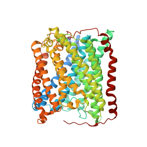
Combined microspectrophotometric and crystallographic examination of chemically reduced and X-ray radiation-reduced forms of cytochrome ba3 oxidase from Thermus thermophilus: structure of the reduced form of the enzyme.

Liu, B.Chen, Y.Doukov, T.Soltis, S.M.Stout, C.D.Fee, J.A.

(2009) Biochemistry 48: 820-826

  • DOI: https://doi.org/10.1021/bi801759a
  • Primary Citation of Related Structures:  
    3EH3, 3EH4, 3EH5

  • PubMed Abstract: 

    Three paths for obtaining crystals of reduced (II-E4Q/I-K258R) cytochrome ba(3) are described, and the structures of these are reported at approximately 2.8-3.0 A resolution. Microspectrophotometry of single crystals of Thermus ba(3) oxidase at 100 K was used to show that crystals of the oxidized enzyme are reduced in an intense X-ray (beam line 7-1 at the Stanford Synchrotron Radiation Laboratory), being nearly complete in 1 min. The previously reported structures of ba(3) (Protein Data Bank entries 1EHK and 1XME ), having a crystallographically detectable water between the Cu(B) and Fe(a3) metals of the dinuclear center, actually represent the X-ray radiation-reduced enzyme. Dithionite-reduced crystals or crystals formed from dithionite-reduced enzyme revealed the absence of the above-mentioned water and an increase in the Cu(B)-Fe(a3) distance of approximately 0.3 A. The new structures are discussed in terms of enzyme function. An unexpected optical absorption envelope at approximately 590 nm is also reported. This spectral feature is tentatively thought to arise from a five-coordinate, low-spin, ferrous heme a(3) that is trapped in the frozen crystals.


  • Organizational Affiliation

    Department of Molecular Biology, The Scripps Research Institute, MB-8, 10550 North Torrey Pines Road, La Jolla, California 92037, USA.


Macromolecules
Find similar proteins by:  (by identity cutoff)  |  3D Structure
Entity ID: 1
MoleculeChains Sequence LengthOrganismDetailsImage
Cytochrome c oxidase subunit 1618Thermus thermophilus HB8Mutation(s): 1 
Gene Names: cbaATTHA1135
EC: 1.9.3.1
Membrane Entity: Yes 
UniProt
Find proteins for Q5SJ79 (Thermus thermophilus (strain ATCC 27634 / DSM 579 / HB8))
Explore Q5SJ79 
Go to UniProtKB:  Q5SJ79
Entity Groups  
Sequence Clusters30% Identity50% Identity70% Identity90% Identity95% Identity100% Identity
UniProt GroupQ5SJ79
Sequence Annotations
Expand
  • Reference Sequence
Find similar proteins by:  (by identity cutoff)  |  3D Structure
Entity ID: 2
MoleculeChains Sequence LengthOrganismDetailsImage
Cytochrome c oxidase subunit 2166Thermus thermophilus HB8Mutation(s): 1 
Gene Names: cbaBctaCTTHA1134
EC: 1.9.3.1
Membrane Entity: Yes 
UniProt
Find proteins for Q5SJ80 (Thermus thermophilus (strain ATCC 27634 / DSM 579 / HB8))
Explore Q5SJ80 
Go to UniProtKB:  Q5SJ80
Entity Groups  
Sequence Clusters30% Identity50% Identity70% Identity90% Identity95% Identity100% Identity
UniProt GroupQ5SJ80
Sequence Annotations
Expand
  • Reference Sequence
Find similar proteins by:  (by identity cutoff)  |  3D Structure
Entity ID: 3
MoleculeChains Sequence LengthOrganismDetailsImage
Cytochrome c oxidase polypeptide 2A33Thermus thermophilus HB8Mutation(s): 0 
Gene Names: cbaDTTHA1133
EC: 1.9.3.1
Membrane Entity: Yes 
UniProt
Find proteins for P82543 (Thermus thermophilus (strain ATCC 27634 / DSM 579 / HB8))
Explore P82543 
Go to UniProtKB:  P82543
Entity Groups  
Sequence Clusters30% Identity50% Identity70% Identity90% Identity95% Identity100% Identity
UniProt GroupP82543
Sequence Annotations
Expand
  • Reference Sequence
Small Molecules
Ligands 5 Unique
IDChains Name / Formula / InChI Key2D Diagram3D Interactions
HAS
Query on HAS

Download Ideal Coordinates CCD File 
F [auth A]HEME-AS
C54 H64 Fe N4 O6
PDYODZVCODUKFH-ZOMLSHGTSA-L
HEM
Query on HEM

Download Ideal Coordinates CCD File 
E [auth A]PROTOPORPHYRIN IX CONTAINING FE
C34 H32 Fe N4 O4
KABFMIBPWCXCRK-RGGAHWMASA-L
BNG
Query on BNG

Download Ideal Coordinates CCD File 
G [auth A],
H [auth A],
I [auth A],
K [auth C]
nonyl beta-D-glucopyranoside
C15 H30 O6
QFAPUKLCALRPLH-UXXRCYHCSA-N
CUA
Query on CUA

Download Ideal Coordinates CCD File 
J [auth B]DINUCLEAR COPPER ION
Cu2
ALKZAGKDWUSJED-UHFFFAOYSA-N
CU1
Query on CU1

Download Ideal Coordinates CCD File 
D [auth A]COPPER (I) ION
Cu
VMQMZMRVKUZKQL-UHFFFAOYSA-N
Experimental Data & Validation

Experimental Data

  • Method: X-RAY DIFFRACTION
  • Resolution: 2.80 Å
  • R-Value Free: 0.257 
  • R-Value Work: 0.197 
  • R-Value Observed: 0.200 
  • Space Group: P 41 21 2
Unit Cell:
Length ( Å )Angle ( ˚ )
a = 115.631α = 90
b = 115.631β = 90
c = 149.169γ = 90
Software Package:
Software NamePurpose
Blu-Icedata collection
PHASERphasing
REFMACrefinement
MOSFLMdata reduction
SCALAdata scaling

Structure Validation

View Full Validation Report


Ligand Structure Quality Assessment 


Entry History 

Deposition Data

Revision History  (Full details and data files)

  • Version 1.0: 2009-04-21
    Type: Initial release
  • Version 1.1: 2011-07-13
    Changes: Advisory, Source and taxonomy, Version format compliance
  • Version 1.2: 2017-10-25
    Changes: Refinement description
  • Version 1.3: 2018-01-24
    Changes: Structure summary
  • Version 1.4: 2020-07-29
    Type: Remediation
    Reason: Carbohydrate remediation
    Changes: Data collection, Database references, Derived calculations, Structure summary
  • Version 1.5: 2021-10-20
    Changes: Database references, Structure summary
  • Version 1.6: 2023-08-30
    Changes: Data collection, Refinement description