3EDJ

Structural base for cyclodextrin hydrolysis


Experimental Data Snapshot

  • Method: X-RAY DIFFRACTION
  • Resolution: 1.69 Å
  • R-Value Free: 0.201 
  • R-Value Work: 0.169 
  • R-Value Observed: 0.169 

wwPDB Validation   3D Report Full Report


This is version 2.1 of the entry. See complete history


Literature

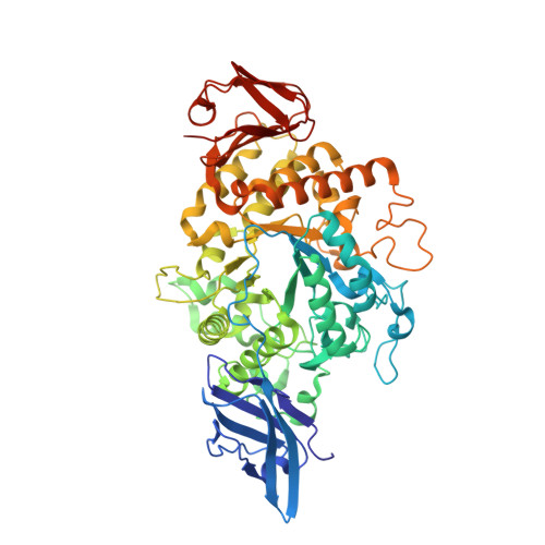
Structural base for enzymatic cyclodextrin hydrolysis

Buedenbender, S.Schulz, G.E.

(2009) J Mol Biol 385: 606-617

  • DOI: https://doi.org/10.1016/j.jmb.2008.10.085
  • Primary Citation of Related Structures:  
    3EDE, 3EDJ

  • PubMed Abstract: 

    Cyclodextrins resist hydrolysis by burying all bridge oxygens at their interior. Still, the rings can be opened by a small group of specialized enzymes, the cyclomaltodextrinases. Among them, the enzyme from Flavobacterium sp. no. 92 was mutated, crystallized and soaked with cyclodextrins, giving rise to four complex structures. One of them showed an alpha-cyclodextrin at the outer rim of the active center pocket. In the other complexes, alpha-, beta-and gamma-cyclodextrins were bound in a competent mode in the active center. The structures suggest that Arg464 functions as a chaperone guiding the substrates from the solvent into the active center. Over the last part of this pathway, the cyclodextrins bump on Phe274, which rotates the glucosyl group at subsite (+1) by about 120 degrees and fixes it in the new conformation. This induced fit was observed with all three major cyclodextrins. It makes the bridging oxygen between subsites (+1) and (-1) available for protonation by Glu340, which starts the hydrolysis. The mechanism resembles a spring-lock. The structural data were supplemented by activity measurements, quantifying the initial ring opening reaction for the major cyclodextrins and the transglucosylation activity for maltotetraose. Further activity data were collected for mutants splitting the tetrameric enzyme into dimers and for active center mutants.


  • Organizational Affiliation

    Institut für Organische Chemie und Biochemie, Albert-Ludwigs-Universität, Albertstr. 21, D-79104 Freiburg im Breisgau, Germany.


Macromolecules
Find similar proteins by:  (by identity cutoff)  |  3D Structure
Entity ID: 1
MoleculeChains Sequence LengthOrganismDetailsImage
Cyclomaltodextrinase
A, B
601Flavobacterium sp. 92Mutation(s): 2 
Gene Names: cdase
EC: 3.2.1.54
UniProt
Find proteins for Q8KKG0 (Flavobacterium sp. 92)
Explore Q8KKG0 
Go to UniProtKB:  Q8KKG0
Entity Groups  
Sequence Clusters30% Identity50% Identity70% Identity90% Identity95% Identity100% Identity
UniProt GroupQ8KKG0
Sequence Annotations
Expand
  • Reference Sequence
Oligosaccharides

Help

Entity ID: 2
MoleculeChains Length2D Diagram Glycosylation3D Interactions
Cycloheptakis-(1-4)-(alpha-D-glucopyranose)
C, D
7N/A
Glycosylation Resources
GlyTouCan:  G01435GL
GlyCosmos:  G01435GL
Biologically Interesting Molecules (External Reference) 1 Unique
Experimental Data & Validation

Experimental Data

  • Method: X-RAY DIFFRACTION
  • Resolution: 1.69 Å
  • R-Value Free: 0.201 
  • R-Value Work: 0.169 
  • R-Value Observed: 0.169 
  • Space Group: P 21 21 2
Unit Cell:
Length ( Å )Angle ( ˚ )
a = 106.464α = 90
b = 111.107β = 90
c = 106.793γ = 90
Software Package:
Software NamePurpose
REFMACrefinement
PDB_EXTRACTdata extraction
MAR345dtbdata collection
XDSdata reduction
XSCALEdata scaling
REFMACphasing

Structure Validation

View Full Validation Report


Entry History 

Deposition Data

Revision History  (Full details and data files)

  • Version 1.0: 2009-03-03
    Type: Initial release
  • Version 1.1: 2011-07-13
    Changes: Non-polymer description, Version format compliance
  • Version 2.0: 2020-07-29
    Type: Remediation
    Reason: Carbohydrate remediation
    Changes: Atomic model, Data collection, Derived calculations, Non-polymer description, Structure summary
  • Version 2.1: 2021-11-10
    Changes: Database references, Structure summary