


Crystal structure of prokaryotic domain of unknown function (DUF849) with a TIM barrel fold (YP_556190.1) from BURKHOLDERIA XENOVORANS LB400 at 1.75 A resolution
Joint Center for Structural Genomics (JCSG)To be published.
Experimental Data Snapshot
wwPDB Validation 3D Report Full Report

Entity ID: 1 | |||||
|---|---|---|---|---|---|
| Molecule | Chains | Sequence Length | Organism | Details | Image |
| uncharacterized protein DUF849 with a TIM barrel fold | 311 | Paraburkholderia xenovorans LB400 | Mutation(s): 0 Gene Names: YP_556190.1, Bxeno_C0912, Bxe_C0966 | 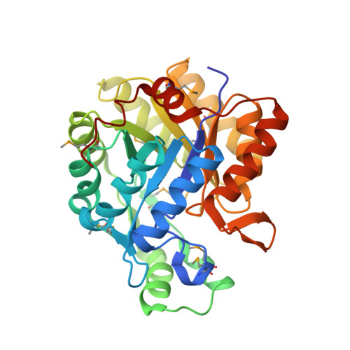 | |
UniProt | |||||
Find proteins for Q13GE9 (Paraburkholderia xenovorans (strain LB400)) Explore Q13GE9 Go to UniProtKB: Q13GE9 | |||||
Entity Groups | |||||
| Sequence Clusters | 30% Identity50% Identity70% Identity90% Identity95% Identity100% Identity | ||||
| UniProt Group | Q13GE9 | ||||
Sequence AnnotationsExpand | |||||
| |||||
| Ligands 4 Unique | |||||
|---|---|---|---|---|---|
| ID | Chains | Name / Formula / InChI Key | 2D Diagram | 3D Interactions | |
| ETX Query on ETX | F [auth A] H [auth B] I [auth B] J [auth B] N [auth C] | 2-ETHOXYETHANOL C4 H10 O2 ZNQVEEAIQZEUHB-UHFFFAOYSA-N |  | ||
| IMD Query on IMD | L [auth B], R [auth C], T [auth D], U [auth D], V [auth D] | IMIDAZOLE C3 H5 N2 RAXXELZNTBOGNW-UHFFFAOYSA-O |  | ||
| ZN Query on ZN | E [auth A], G [auth B], M [auth C], S [auth D] | ZINC ION Zn PTFCDOFLOPIGGS-UHFFFAOYSA-N |  | ||
| ACT Query on ACT | K [auth B], Q [auth C] | ACETATE ION C2 H3 O2 QTBSBXVTEAMEQO-UHFFFAOYSA-M |  | ||
| Modified Residues 1 Unique | |||||
|---|---|---|---|---|---|
| ID | Chains | Type | Formula | 2D Diagram | Parent |
| MSE Query on MSE | A, B, C, D | L-PEPTIDE LINKING | C5 H11 N O2 Se |  | MET |
| Length ( Å ) | Angle ( ˚ ) |
|---|---|
| a = 59.495 | α = 69.99 |
| b = 68.275 | β = 88.07 |
| c = 83.952 | γ = 72.84 |
| Software Name | Purpose |
|---|---|
| REFMAC | refinement |
| PHENIX | refinement |
| SHELX | phasing |
| MolProbity | model building |
| SCALA | data scaling |
| PDB_EXTRACT | data extraction |
| MOSFLM | data reduction |
| SHELXD | phasing |
| autoSHARP | phasing |
RCSB PDB (citation) is hosted by
RCSB PDB is a member of the

