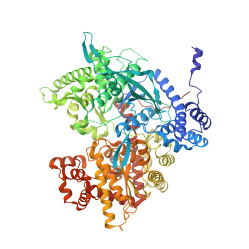
3E3L

The R-state Glycogen Phosphorylase


Experimental Data Snapshot

  • Method: X-RAY DIFFRACTION
  • Resolution: 2.59 Å
  • R-Value Free: 0.266 
  • R-Value Work: 0.207 
  • R-Value Observed: 0.210 

wwPDB Validation   3D Report Full Report


This is version 1.5 of the entry. See complete history


Literature

Glycogen phosphorylase revisited: extending the resolution of the R- and T-state structures of the free enzyme and in complex with allosteric activators.

Leonidas, D.D.Zographos, S.E.Tsitsanou, K.E.Skamnaki, V.T.Stravodimos, G.Kyriakis, E.

(2021) Acta Crystallogr F Struct Biol Commun 77: 303-311

  • DOI: https://doi.org/10.1107/S2053230X21008542
  • Primary Citation of Related Structures:  
    3E3L, 3E3N, 3E3O, 7P7D

  • PubMed Abstract: 

    The crystal structures of free T-state and R-state glycogen phosphorylase (GP) and of R-state GP in complex with the allosteric activators IMP and AMP are reported at improved resolution. GP is a validated pharmaceutical target for the development of antihyperglycaemic agents, and the reported structures may have a significant impact on structure-based drug-design efforts. Comparisons with previously reported structures at lower resolution reveal the detailed conformation of important structural features in the allosteric transition of GP from the T-state to the R-state. The conformation of the N-terminal segment (residues 7-17), the position of which was not located in previous T-state structures, was revealed to form an α-helix (now termed α0). The conformation of this segment (which contains Ser14, phosphorylation of which leads to the activation of GP) is significantly different between the T-state and the R-state, pointing in opposite directions. In the T-state it is packed between helices α4 and α16 (residues 104-115 and 497-508, respectively), while in the R-state it is packed against helix α1 (residues 22'-38') and towards the loop connecting helices α4' and α5' of the neighbouring subunit. The allosteric binding site where AMP and IMP bind is formed by the ordering of a loop (residues 313-326) which is disordered in the free structure, and adopts a conformation dictated mainly by the type of nucleotide that binds at this site.


  • Organizational Affiliation

    Department of Biochemistry and Biotechnology, University of Thessaly, Biopolis, 41500 Larissa, Greece.


Macromolecules
Find similar proteins by:  (by identity cutoff)  |  3D Structure
Entity ID: 1
MoleculeChains Sequence LengthOrganismDetailsImage
Glycogen phosphorylase, muscle form
A, B, C, D
842Oryctolagus cuniculusMutation(s): 0 
EC: 2.4.1.1
UniProt
Find proteins for P00489 (Oryctolagus cuniculus)
Explore P00489 
Go to UniProtKB:  P00489
Entity Groups  
Sequence Clusters30% Identity50% Identity70% Identity90% Identity95% Identity100% Identity
UniProt GroupP00489
Sequence Annotations
Expand
  • Reference Sequence
Small Molecules
Ligands 1 Unique
IDChains Name / Formula / InChI Key2D Diagram3D Interactions
SO4
Query on SO4

Download Ideal Coordinates CCD File 
E [auth A]
F [auth A]
G [auth A]
H [auth B]
I [auth B]
E [auth A],
F [auth A],
G [auth A],
H [auth B],
I [auth B],
J [auth B],
K [auth C],
L [auth C],
M [auth D],
N [auth D],
O [auth D],
P [auth D]
SULFATE ION
O4 S
QAOWNCQODCNURD-UHFFFAOYSA-L
Modified Residues  1 Unique
IDChains TypeFormula2D DiagramParent
LLP
Query on LLP
A, B, C, D
L-PEPTIDE LINKINGC14 H22 N3 O7 PLYS
Experimental Data & Validation

Experimental Data

  • Method: X-RAY DIFFRACTION
  • Resolution: 2.59 Å
  • R-Value Free: 0.266 
  • R-Value Work: 0.207 
  • R-Value Observed: 0.210 
  • Space Group: P 1 21 1
Unit Cell:
Length ( Å )Angle ( ˚ )
a = 118.895α = 90
b = 189.92β = 109.27
c = 88.16γ = 90
Software Package:
Software NamePurpose
REFMACrefinement
MAR345data collection
DENZOdata reduction
SCALEPACKdata scaling
REFMACphasing

Structure Validation

View Full Validation Report


Entry History 

Deposition Data

Revision History  (Full details and data files)

  • Version 1.0: 2009-02-10
    Type: Initial release
  • Version 1.1: 2011-07-13
    Changes: Advisory, Version format compliance
  • Version 1.2: 2017-10-25
    Changes: Refinement description
  • Version 1.3: 2021-09-15
    Changes: Database references, Derived calculations
  • Version 1.4: 2023-11-01
    Changes: Data collection, Refinement description
  • Version 1.5: 2023-11-15
    Changes: Data collection