3DYB

proteinase K- digalacturonic acid complex


Experimental Data Snapshot

  • Method: X-RAY DIFFRACTION
  • Resolution: 1.32 Å
  • R-Value Free: 0.207 
  • R-Value Work: 0.158 
  • R-Value Observed: 0.160 

wwPDB Validation   3D Report Full Report


This is version 2.2 of the entry. See complete history


Literature

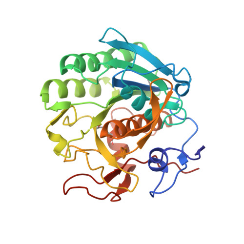
High-resolution structure of proteinase K cocrystallized with digalacturonic acid.

Larson, S.B.Day, J.S.Nguyen, C.Cudney, R.McPherson, A.

(2009) Acta Crystallogr Sect F Struct Biol Cryst Commun 65: 192-198

  • DOI: https://doi.org/10.1107/S1744309109002218
  • Primary Citation of Related Structures:  
    3DYB

  • PubMed Abstract: 

    Proteinase K, a subtilisin-like fungal protease, was crystallized from a cocktail of small molecules containing digalacturonic acid (DGA). The crystal structure was determined to 1.32 A resolution and refined to an R factor of 0.158. The final model contained, beside the protein, two calcium ions, 379 water molecules, a molecule of DGA and a partially occupied HEPES molecule. The DGA molecule has one sugar moiety disposed exactly on a crystallographic twofold axis; the second ring was not observed. The DGA molecule is bound to two protein molecules across the twofold axis through hydrogen-bonding networks involving Ser150 and water molecules. One of the calcium-ion sites has not been reported previously. This study further illustrates the involvement of small molecules in the crystallization of macromolecules through their ability to form intermolecular lattice interactions.


  • Organizational Affiliation

    Department of Molecular Biology and Biochemistry, The University of California, Irvine, 92697-3900, USA.


Macromolecules
Find similar proteins by:  (by identity cutoff)  |  3D Structure
Entity ID: 1
MoleculeChains Sequence LengthOrganismDetailsImage
Proteinase K279Parengyodontium albumMutation(s): 0 
EC: 3.4.21.64
UniProt
Find proteins for P06873 (Parengyodontium album)
Explore P06873 
Go to UniProtKB:  P06873
Entity Groups  
Sequence Clusters30% Identity50% Identity70% Identity90% Identity95% Identity100% Identity
UniProt GroupP06873
Sequence Annotations
Expand
  • Reference Sequence
Oligosaccharides

Help

Entity ID: 2
MoleculeChains Length2D Diagram Glycosylation3D Interactions
alpha-D-galactopyranuronic acid-(1-4)-alpha-D-galactopyranuronic acid
B
2N/A
Glycosylation Resources
GlyTouCan:  G65215OP
GlyCosmos:  G65215OP
Experimental Data & Validation

Experimental Data

  • Method: X-RAY DIFFRACTION
  • Resolution: 1.32 Å
  • R-Value Free: 0.207 
  • R-Value Work: 0.158 
  • R-Value Observed: 0.160 
  • Space Group: P 43 21 2
Unit Cell:
Length ( Å )Angle ( ˚ )
a = 67.72α = 90
b = 67.72β = 90
c = 101.89γ = 90
Software Package:
Software NamePurpose
REFMACrefinement
d*TREKdata reduction
d*TREKdata scaling

Structure Validation

View Full Validation Report


Entry History 

Deposition Data

Revision History  (Full details and data files)

  • Version 1.0: 2008-10-07
    Type: Initial release
  • Version 1.1: 2011-07-13
    Changes: Atomic model, Version format compliance
  • Version 1.2: 2017-10-25
    Changes: Advisory, Refinement description
  • Version 2.0: 2020-07-29
    Type: Remediation
    Reason: Carbohydrate remediation
    Changes: Advisory, Atomic model, Data collection, Derived calculations, Non-polymer description, Structure summary
  • Version 2.1: 2021-11-17
    Changes: Advisory, Database references, Structure summary
  • Version 2.2: 2023-08-30
    Changes: Data collection, Refinement description