3CVR

Crystal structure of the full length IpaH3


Experimental Data Snapshot

  • Method: X-RAY DIFFRACTION
  • Resolution: 2.80 Å
  • R-Value Free: 0.277 
  • R-Value Work: 0.251 
  • R-Value Observed: 0.252 

wwPDB Validation   3D Report Full Report


This is version 1.1 of the entry. See complete history


Literature

Structure of a Shigella effector reveals a new class of ubiquitin ligases

Zhu, Y.Li, H.Hu, L.Wang, J.Zhou, Y.Pang, Z.Liu, L.Shao, F.

(2008) Nat Struct Mol Biol 15: 1302-1308

  • DOI: https://doi.org/10.1038/nsmb.1517

  • PubMed Abstract: 

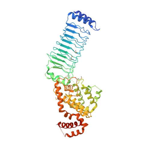
    Bacterial pathogens have evolved effector proteins with ubiquitin E3 ligase activities through structural mimicking. Here we report the crystal structure of the Shigella flexneri type III effector IpaH3, a member of the leucine-rich repeat (LRR)-containing bacterial E3 family. The LRR domain is structurally similar to Yersinia pestis YopM and potentially binds to substrates. The structure of the C-terminal E3 domain differs from the typical RING- and HECT-type E3s. IpaH3 synthesizes a Lys48-linked ubiquitin chain, and the reaction requires noncovalent binding between ubiquitin and a specific E2, UbcH5. Free ubiquitin serves as an acceptor for IpaH3-catalyzed ubiquitin transfer. Cys363 within a conserved CXD motif acts as a nucleophile to catalyze ubiquitin transfer through a transthiolation reaction. The D365N mutant is devoid of E3 activities but turns into a potent ubiquitin-E2 thioesterase. Our analysis establishes a structurally and mechanistically distinct class of ubiquitin ligases found exclusively in pathogenic or symbiotic bacteria.


  • Organizational Affiliation

    National Institute of Biological Sciences, 7# Science Park Road, Zhongguancun Life Science Park, Beijing 102206, China.


Macromolecules
Find similar proteins by:  (by identity cutoff)  |  3D Structure
Entity ID: 1
MoleculeChains Sequence LengthOrganismDetailsImage
Invasion plasmid antigen571Shigella flexneri 2a str. 301Mutation(s): 0 
Gene Names: ipaH_3
EC: 6.3.2
UniProt
Find proteins for Q83RJ4 (Shigella flexneri)
Explore Q83RJ4 
Go to UniProtKB:  Q83RJ4
Entity Groups  
Sequence Clusters30% Identity50% Identity70% Identity90% Identity95% Identity100% Identity
UniProt GroupQ83RJ4
Sequence Annotations
Expand
  • Reference Sequence
Small Molecules
Modified Residues  1 Unique
IDChains TypeFormula2D DiagramParent
MSE
Query on MSE
A
L-PEPTIDE LINKINGC5 H11 N O2 SeMET
Experimental Data & Validation

Experimental Data

  • Method: X-RAY DIFFRACTION
  • Resolution: 2.80 Å
  • R-Value Free: 0.277 
  • R-Value Work: 0.251 
  • R-Value Observed: 0.252 
  • Space Group: P 41 21 2
Unit Cell:
Length ( Å )Angle ( ˚ )
a = 154.19α = 90
b = 154.19β = 90
c = 85.87γ = 90
Software Package:
Software NamePurpose
SCALAdata processing
SOLVEphasing
CNSrefinement
PDB_EXTRACTdata extraction
ADSCdata collection
MOSFLMdata reduction
SCALAdata scaling
REFMACrefinement

Structure Validation

View Full Validation Report


Entry History 

Deposition Data

Revision History  (Full details and data files)

  • Version 1.0: 2008-11-11
    Type: Initial release
  • Version 1.1: 2011-07-13
    Changes: Advisory, Version format compliance