3CQ5

Histidinol-phosphate aminotransferase from Corynebacterium glutamicum in complex with PMP


Experimental Data Snapshot

  • Method: X-RAY DIFFRACTION
  • Resolution: 1.80 Å
  • R-Value Free: 0.219 
  • R-Value Work: 0.192 
  • R-Value Observed: 0.193 

wwPDB Validation   3D Report Full Report


Ligand Structure Quality Assessment 


This is version 1.2 of the entry. See complete history


Literature

Insights into the structural basis of substrate recognition by histidinol-phosphate aminotransferase from Corynebacterium glutamicum

Marienhagen, J.Sandalova, T.Sahm, H.Eggeling, L.Schneider, G.

(2008) Acta Crystallogr D Biol Crystallogr 64: 675-685

  • DOI: https://doi.org/10.1107/S0907444908009438
  • Primary Citation of Related Structures:  
    3CQ4, 3CQ5, 3CQ6

  • PubMed Abstract: 

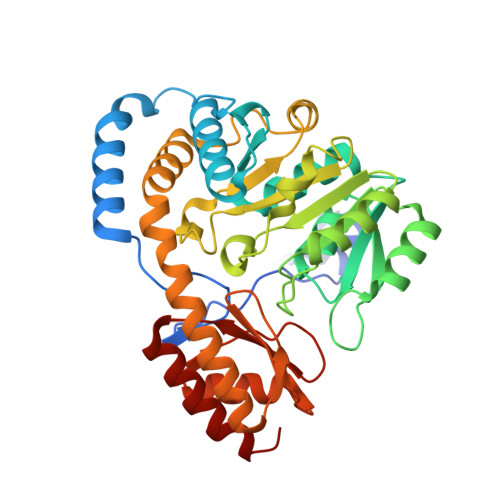
    Histidinol-phosphate aminotransferase (HisC) is a pyridoxal 5'-phosphate-dependent enzyme that catalyzes the reversible transamination reaction between histidinol phosphate (His-P) and 2-oxoglutarate (O-Glu). The crystal structures of apo histidinol-phosphate aminotransferase from Corynebacterium glutamicum, of the internal PLP aldimine adduct and of a pyridoxamine 5-phosphate-enzyme complex were determined at resolutions of 2.2, 2.1 and 1.8 A, respectively. Residues important for substrate specificity were identified by modelling His-P into the active site and comparison with crystal structures of HisC from Thermotoga maritima and Escherichia coli. Four of the residues lining the substrate-binding pocket were studied by site-directed mutagenesis. Kinetic analysis of the Tyr21Phe mutant suggested that the hydrogen bond between the side chain of this residue and the phosphate group of His-P is important for recognition of the natural substrate and discrimination against other potential amino donors such as phenylalanine and leucine. The mutagenesis studies further indicated that residue Asn99 does not contribute to the specific recognition of the amino-acid donor, but may be involved in binding of the phosphate group of pyridoxal 5'-phosphate. The conserved residues Tyr123 and Tyr257 interact with the substrate through van der Waals interactions and their potential for hydrogen-bonding interactions is not utilized in substrate recognition, as the corresponding phenylalanine mutants show only a moderate effect on the catalytic efficiency kcat/Km.


  • Organizational Affiliation

    Institute for Biotechnology, Research Center Juelich, 52425 Juelich, Germany.


Macromolecules
Find similar proteins by:  (by identity cutoff)  |  3D Structure
Entity ID: 1
MoleculeChains Sequence LengthOrganismDetailsImage
Histidinol-phosphate aminotransferase
A, B, C
369Corynebacterium glutamicumMutation(s): 0 
Gene Names: HisC
EC: 2.6.1.9
UniProt
Find proteins for Q9KJU4 (Corynebacterium glutamicum (strain ATCC 13032 / DSM 20300 / BCRC 11384 / JCM 1318 / LMG 3730 / NCIMB 10025))
Explore Q9KJU4 
Go to UniProtKB:  Q9KJU4
Entity Groups  
Sequence Clusters30% Identity50% Identity70% Identity90% Identity95% Identity100% Identity
UniProt GroupQ9KJU4
Sequence Annotations
Expand
  • Reference Sequence
Small Molecules
Ligands 3 Unique
IDChains Name / Formula / InChI Key2D Diagram3D Interactions
PMP
Query on PMP

Download Ideal Coordinates CCD File 
H [auth A],
K [auth B],
O [auth C]
4'-DEOXY-4'-AMINOPYRIDOXAL-5'-PHOSPHATE
C8 H13 N2 O5 P
ZMJGSOSNSPKHNH-UHFFFAOYSA-N
144
Query on 144

Download Ideal Coordinates CCD File 
G [auth A],
I [auth A],
N [auth C]
TRIS-HYDROXYMETHYL-METHYL-AMMONIUM
C4 H12 N O3
DRDCQJADRSJFFD-UHFFFAOYSA-N
SO4
Query on SO4

Download Ideal Coordinates CCD File 
D [auth A]
E [auth A]
F [auth A]
J [auth B]
L [auth C]
D [auth A],
E [auth A],
F [auth A],
J [auth B],
L [auth C],
M [auth C]
SULFATE ION
O4 S
QAOWNCQODCNURD-UHFFFAOYSA-L
Experimental Data & Validation

Experimental Data

  • Method: X-RAY DIFFRACTION
  • Resolution: 1.80 Å
  • R-Value Free: 0.219 
  • R-Value Work: 0.192 
  • R-Value Observed: 0.193 
  • Space Group: C 1 2 1
Unit Cell:
Length ( Å )Angle ( ˚ )
a = 195.273α = 90
b = 85.531β = 93.65
c = 89.431γ = 90
Software Package:
Software NamePurpose
REFMACrefinement
DNAdata collection
MOSFLMdata reduction
SCALAdata scaling
MOLREPphasing

Structure Validation

View Full Validation Report


Ligand Structure Quality Assessment 


Entry History 

Deposition Data

Revision History  (Full details and data files)

  • Version 1.0: 2008-07-01
    Type: Initial release
  • Version 1.1: 2011-07-13
    Changes: Version format compliance
  • Version 1.2: 2024-03-13
    Changes: Advisory, Data collection, Database references, Derived calculations, Refinement description