3CIQ

A regulatable switch mediates self-association in an immunoglobulin fold


Experimental Data Snapshot

  • Method: X-RAY DIFFRACTION
  • Resolution: 2.90 Å
  • R-Value Free: 0.264 
  • R-Value Work: 0.225 
  • R-Value Observed: 0.227 

wwPDB Validation   3D Report Full Report


This is version 1.2 of the entry. See complete history


Literature

A regulatable switch mediates self-association in an immunoglobulin fold.

Calabrese, M.F.Eakin, C.M.Wang, J.M.Miranker, A.D.

(2008) Nat Struct Mol Biol 15: 965-971

  • DOI: https://doi.org/10.1038/nsmb.1483
  • Primary Citation of Related Structures:  
    3CIQ

  • PubMed Abstract: 

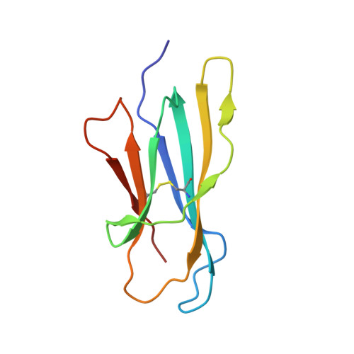
    Beta-2 microglobulin (beta2m) is a globular protein that self-associates into fibrillar amyloid deposits in patients undergoing hemodialysis therapy. Formation of these beta-sheet-rich assemblies is a fundamental property of polypeptides that can be triggered by diverse conditions. For beta2m, oligomerization into pre-amyloidogenic states occurs in specific response to coordination by Cu2+. Here we report the basis for this self-association at atomic resolution. Metal is not a direct participant in the molecular interface. Rather, binding results in distal alterations enabling the formation of two new surfaces. These interact to form a closed hexameric species. The origins of this include isomerization of a buried and conserved cis-proline previously implicated in the beta2m aggregation pathway. The consequences of this isomerization are evident and reveal a molecular basis for the conversion of this robust monomeric protein into an amyloid-competent state.


  • Organizational Affiliation

    Department of Molecular Biophysics and Biochemistry, Yale University, 260 Whitney Avenue, New Haven, Connecticut 06520-8114, USA.


Macromolecules
Find similar proteins by:  (by identity cutoff)  |  3D Structure
Entity ID: 1
MoleculeChains Sequence LengthOrganismDetailsImage
Beta-2-microglobulin
A, B, C, D, E
A, B, C, D, E, F, G, H, I, J, K, L
100Homo sapiensMutation(s): 1 
Gene Names: B2M
UniProt & NIH Common Fund Data Resources
Find proteins for P61769 (Homo sapiens)
Explore P61769 
Go to UniProtKB:  P61769
PHAROS:  P61769
GTEx:  ENSG00000166710 
Entity Groups  
Sequence Clusters30% Identity50% Identity70% Identity90% Identity95% Identity100% Identity
UniProt GroupP61769
Sequence Annotations
Expand
  • Reference Sequence
Small Molecules
Ligands 2 Unique
IDChains Name / Formula / InChI Key2D Diagram3D Interactions
TLA
Query on TLA

Download Ideal Coordinates CCD File 
T [auth G]L(+)-TARTARIC ACID
C4 H6 O6
FEWJPZIEWOKRBE-JCYAYHJZSA-N
CU
Query on CU

Download Ideal Coordinates CCD File 
M [auth A]
N [auth B]
O [auth C]
P [auth D]
Q [auth E]
M [auth A],
N [auth B],
O [auth C],
P [auth D],
Q [auth E],
R [auth F],
S [auth G],
U [auth H],
V [auth I],
W [auth J],
X [auth K],
Y [auth L]
COPPER (II) ION
Cu
JPVYNHNXODAKFH-UHFFFAOYSA-N
Experimental Data & Validation

Experimental Data

  • Method: X-RAY DIFFRACTION
  • Resolution: 2.90 Å
  • R-Value Free: 0.264 
  • R-Value Work: 0.225 
  • R-Value Observed: 0.227 
  • Space Group: P 1
Unit Cell:
Length ( Å )Angle ( ˚ )
a = 67.594α = 104.38
b = 68.047β = 94.15
c = 96.218γ = 117.77
Software Package:
Software NamePurpose
REFMACrefinement
HKL-2000data reduction
HKL-2000data scaling
PHASERphasing

Structure Validation

View Full Validation Report


Entry History 

Deposition Data

Revision History  (Full details and data files)

  • Version 1.0: 2008-09-02
    Type: Initial release
  • Version 1.1: 2011-07-13
    Changes: Advisory, Version format compliance
  • Version 1.2: 2021-10-20
    Changes: Database references, Derived calculations