3C39

Crystal Structure of human phosphoglycerate kinase bound to 3-phosphoglycerate


Experimental Data Snapshot

  • Method: X-RAY DIFFRACTION
  • Resolution: 1.85 Å
  • R-Value Free: 0.273 
  • R-Value Work: 0.191 
  • R-Value Observed: 0.194 

wwPDB Validation   3D Report Full Report


Ligand Structure Quality Assessment 


This is version 1.2 of the entry. See complete history


Literature

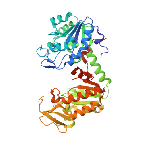
Molecular basis for the lack of enantioselectivity of human 3-phosphoglycerate kinase

Gondeau, C.Chaloin, L.Lallemand, P.Roy, B.Perigaud, C.Barman, T.Varga, A.Vas, M.Lionne, C.Arold, S.T.

(2008) Nucleic Acids Res 36: 3620-3629

  • DOI: https://doi.org/10.1093/nar/gkn212
  • Primary Citation of Related Structures:  
    2ZGV, 3C39, 3C3A, 3C3B, 3C3C

  • PubMed Abstract: 

    Non-natural L-nucleoside analogues are increasingly used as therapeutic agents to treat cancer and viral infections. To be active, L-nucleosides need to be phosphorylated to their respective triphosphate metabolites. This stepwise phosphorylation relies on human enzymes capable of processing L-nucleoside enantiomers. We used crystallographic analysis to reveal the molecular basis for the low enantioselectivity and the broad specificity of human 3-phosphoglycerate kinase (hPGK), an enzyme responsible for the last step of phosphorylation of many nucleotide derivatives. Based on structures of hPGK in the absence of nucleotides, and bound to L and d forms of MgADP and MgCDP, we show that a non-specific hydrophobic clamp to the nucleotide base, as well as a water-filled cavity behind it, allows high flexibility in the interaction between PGK and the bases. This, combined with the dispensability of hydrogen bonds to the sugar moiety, and ionic interactions with the phosphate groups, results in the positioning of different nucleotides so to expose their diphosphate group in a position competent for catalysis. Since the third phosphorylation step is often rate limiting, our results are expected to alleviate in silico tailoring of L-type prodrugs to assure their efficient metabolic processing.


  • Organizational Affiliation

    Centre d'études d'agents Pathogènes et Biotechnologies pour la Santé, UMR 5236, CNRS-Universités Montpellier 1 et 2, Institut de Biologie, 4 bd Henri IV, CS69033, 34965 Montpellier cedex 2, France.


Macromolecules
Find similar proteins by:  (by identity cutoff)  |  3D Structure
Entity ID: 1
MoleculeChains Sequence LengthOrganismDetailsImage
Phosphoglycerate kinase 1
A, B
420Homo sapiensMutation(s): 0 
Gene Names: PGK1
EC: 2.7.2.3
UniProt & NIH Common Fund Data Resources
Find proteins for P00558 (Homo sapiens)
Explore P00558 
Go to UniProtKB:  P00558
PHAROS:  P00558
GTEx:  ENSG00000102144 
Entity Groups  
Sequence Clusters30% Identity50% Identity70% Identity90% Identity95% Identity100% Identity
UniProt GroupP00558
Sequence Annotations
Expand
  • Reference Sequence
Small Molecules
Binding Affinity Annotations 
IDSourceBinding Affinity
3PG BindingDB:  3C39 Kd: 1.09e+5 (nM) from 1 assay(s)
PDBBind:  3C39 Kd: 1.09e+5 (nM) from 1 assay(s)
Experimental Data & Validation

Experimental Data

  • Method: X-RAY DIFFRACTION
  • Resolution: 1.85 Å
  • R-Value Free: 0.273 
  • R-Value Work: 0.191 
  • R-Value Observed: 0.194 
  • Space Group: P 1
Unit Cell:
Length ( Å )Angle ( ˚ )
a = 35.616α = 79.18
b = 55.79β = 84.33
c = 93.23γ = 83.23
Software Package:
Software NamePurpose
REFMACrefinement
ADSCdata collection
MOSFLMdata reduction
SCALAdata scaling
MOLREPphasing

Structure Validation

View Full Validation Report


Ligand Structure Quality Assessment 


Entry History 

Deposition Data

Revision History  (Full details and data files)

  • Version 1.0: 2008-07-01
    Type: Initial release
  • Version 1.1: 2011-07-13
    Changes: Version format compliance
  • Version 1.2: 2023-11-01
    Changes: Data collection, Database references, Derived calculations, Refinement description