3BOF

Cobalamin-dependent methionine synthase (1-566) from Thermotoga maritima complexed with Zn2+ and Homocysteine


Experimental Data Snapshot

  • Method: X-RAY DIFFRACTION
  • Resolution: 1.70 Å
  • R-Value Free: 0.222 
  • R-Value Work: 0.195 
  • R-Value Observed: 0.196 

wwPDB Validation   3D Report Full Report


This is version 1.3 of the entry. See complete history


Literature

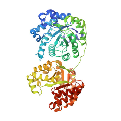
Metal active site elasticity linked to activation of homocysteine in methionine synthases.

Koutmos, M.Pejchal, R.Bomer, T.M.Matthews, R.G.Smith, J.L.Ludwig, M.L.

(2008) Proc Natl Acad Sci U S A 105: 3286-3291

  • DOI: https://doi.org/10.1073/pnas.0709960105
  • Primary Citation of Related Structures:  
    3BOF, 3BOL, 3BQ5, 3BQ6

  • PubMed Abstract: 

    Enzymes possessing catalytic zinc centers perform a variety of fundamental processes in nature, including methyl transfer to thiols. Cobalamin-independent (MetE) and cobalamin-dependent (MetH) methionine synthases are two such enzyme families. Although they perform the same net reaction, transfer of a methyl group from methyltetrahydrofolate to homocysteine (Hcy) to form methionine, they display markedly different catalytic strategies, modular organization, and active site zinc centers. Here we report crystal structures of zinc-replete MetE and MetH, both in the presence and absence of Hcy. Structural investigation of the catalytic zinc sites of these two methyltransferases reveals an unexpected inversion of zinc geometry upon binding of Hcy and displacement of an endogenous ligand in both enzymes. In both cases a significant movement of the zinc relative to the protein scaffold accompanies inversion. These structures provide new information on the activation of thiols by zinc-containing enzymes and have led us to propose a paradigm for the mechanism of action of the catalytic zinc sites in these and related methyltransferases. Specifically, zinc is mobile in the active sites of MetE and MetH, and its dynamic nature helps facilitate the active site conformational changes necessary for thiol activation and methyl transfer.


  • Organizational Affiliation

    Biophysics Research Division, Life Sciences Institute, and Department of Biological Chemistry, University of Michigan, Ann Arbor, MI 48109, USA. mkoutmos@umich.edu


Macromolecules
Find similar proteins by:  (by identity cutoff)  |  3D Structure
Entity ID: 1
MoleculeChains Sequence LengthOrganismDetailsImage
5-methyltetrahydrofolate S-homocysteine methyltransferase
A, B
566Thermotoga maritimaMutation(s): 0 
EC: 2.1.1.13
UniProt
Find proteins for Q9WYA5 (Thermotoga maritima (strain ATCC 43589 / DSM 3109 / JCM 10099 / NBRC 100826 / MSB8))
Explore Q9WYA5 
Go to UniProtKB:  Q9WYA5
Entity Groups  
Sequence Clusters30% Identity50% Identity70% Identity90% Identity95% Identity100% Identity
UniProt GroupQ9WYA5
Sequence Annotations
Expand
  • Reference Sequence
Small Molecules
Ligands 4 Unique
IDChains Name / Formula / InChI Key2D Diagram3D Interactions
HCS
Query on HCS

Download Ideal Coordinates CCD File 
F [auth A],
I [auth B]
2-AMINO-4-MERCAPTO-BUTYRIC ACID
C4 H9 N O2 S
FFFHZYDWPBMWHY-VKHMYHEASA-N
YT3
Query on YT3

Download Ideal Coordinates CCD File 
E [auth A]YTTRIUM (III) ION
Y
GRTBAGCGDOYUBE-UHFFFAOYSA-N
ZN
Query on ZN

Download Ideal Coordinates CCD File 
C [auth A],
G [auth B]
ZINC ION
Zn
PTFCDOFLOPIGGS-UHFFFAOYSA-N
K
Query on K

Download Ideal Coordinates CCD File 
D [auth A],
H [auth B]
POTASSIUM ION
K
NPYPAHLBTDXSSS-UHFFFAOYSA-N
Experimental Data & Validation

Experimental Data

  • Method: X-RAY DIFFRACTION
  • Resolution: 1.70 Å
  • R-Value Free: 0.222 
  • R-Value Work: 0.195 
  • R-Value Observed: 0.196 
  • Space Group: P 1 21 1
Unit Cell:
Length ( Å )Angle ( ˚ )
a = 59.067α = 90
b = 86.308β = 100.03
c = 125.879γ = 90
Software Package:
Software NamePurpose
XSCALEdata scaling
EPMRphasing
REFMACrefinement
PDB_EXTRACTdata extraction
Blu-Icedata collection
XDSdata reduction
CNSrefinement

Structure Validation

View Full Validation Report


Entry History 

Deposition Data

Revision History  (Full details and data files)

  • Version 1.0: 2008-03-11
    Type: Initial release
  • Version 1.1: 2011-07-13
    Changes: Version format compliance
  • Version 1.2: 2024-02-21
    Changes: Data collection, Database references, Derived calculations
  • Version 1.3: 2024-04-03
    Changes: Structure summary