3BLU

crystal structure YopH complexed with inhibitor PVS


Experimental Data Snapshot

  • Method: X-RAY DIFFRACTION
  • Resolution: 2.00 Å
  • R-Value Free: 0.253 
  • R-Value Work: 0.205 

wwPDB Validation   3D Report Full Report


Ligand Structure Quality Assessment 


This is version 1.4 of the entry. See complete history


Literature

Aryl vinyl sulfonates and sulfones as active site-directed and mechanism-based probes for protein tyrosine phosphatases

Liu, S.Zhou, B.Yang, H.He, Y.Jiang, Z.X.Kumar, S.Wu, L.Zhang, Z.Y.

(2008) J Am Chem Soc 130: 8251-8260

  • DOI: https://doi.org/10.1021/ja711125p
  • Primary Citation of Related Structures:  
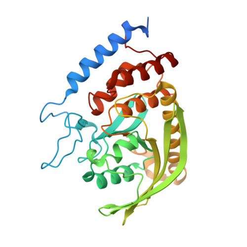
    3BLT, 3BLU, 3BM8

  • PubMed Abstract: 

    Protein tyrosine phosphatases (PTPs) play key roles in the regulation of normal and pathological processes ranging from cell proliferation, differentiation, metabolism, and survival to many human diseases including cancer and diabetes. Functional studies of PTP can be greatly facilitated by small molecule probes that covalently label the active site of a PTP through an activity-dependent chemical reaction. In this article, we characterize phenyl vinyl sulfonate (PVSN) and phenyl vinyl sulfone (PVS) as a new class of mechanism-based PTP probes. PVSN and PVS inactivate a broad range of PTPs in a time- and concentration-dependent fashion. The PVSN- and PVS-mediated PTP inactivation is active site-directed and irreversible, resulting from a Michael addition of the active-site Cys Sgamma onto the terminal carbon of the vinyl group. Structural and mechanistic analyses reveal the molecular basis for the preference of PVSN/PVS toward the PTPs, which lies in the ability of PVSN and PVS to engage the conserved structural and catalytic machinery of the PTP active site. In contrast to early alpha-bromobenzyl phosphonate-based probes, PVSN and PVS are resistant to solvolysis and are cell-permeable and thus hold promise for in vivo applications. Collectively, these properties bode well for the development of aryl vinyl sulfonate/sulfone-based PTP probes to interrogate PTP activity in complex proteomes.


  • Organizational Affiliation

    Department of Biochemistry and Molecular Biology, Indiana University School of Medicine, 635 Barnhill Drive, Indianapolis, Indiana 46202, USA.


Macromolecules
Find similar proteins by:  (by identity cutoff)  |  3D Structure
Entity ID: 1
MoleculeChains Sequence LengthOrganismDetailsImage
Tyrosine-protein phosphatase yopH305Yersinia enterocoliticaMutation(s): 1 
Gene Names: yopHyop51
EC: 3.1.3.48
UniProt
Find proteins for P15273 (Yersinia enterocolitica)
Explore P15273 
Go to UniProtKB:  P15273
Entity Groups  
Sequence Clusters30% Identity50% Identity70% Identity90% Identity95% Identity100% Identity
UniProt GroupP15273
Sequence Annotations
Expand
  • Reference Sequence
Small Molecules
Ligands 1 Unique
IDChains Name / Formula / InChI Key2D Diagram3D Interactions
PVS
Query on PVS

Download Ideal Coordinates CCD File 
B [auth A](ethenylsulfonyl)benzene
C8 H8 O2 S
UJTPZISIAWDGFF-UHFFFAOYSA-N
Binding Affinity Annotations 
IDSourceBinding Affinity
PVS PDBBind:  3BLU Ki: 3.50e+5 (nM) from 1 assay(s)
Experimental Data & Validation

Experimental Data

  • Method: X-RAY DIFFRACTION
  • Resolution: 2.00 Å
  • R-Value Free: 0.253 
  • R-Value Work: 0.205 
  • Space Group: P 21 21 21
Unit Cell:
Length ( Å )Angle ( ˚ )
a = 48.849α = 90
b = 54.092β = 90
c = 96.926γ = 90
Software Package:
Software NamePurpose
DENZOdata reduction
CNSrefinement
HKL-2000data reduction
HKL-2000data scaling
CNSphasing

Structure Validation

View Full Validation Report


Ligand Structure Quality Assessment 


Entry History 

Deposition Data

Revision History  (Full details and data files)

  • Version 1.0: 2008-10-21
    Type: Initial release
  • Version 1.1: 2011-07-13
    Changes: Version format compliance
  • Version 1.2: 2018-01-24
    Changes: Structure summary
  • Version 1.3: 2021-10-20
    Changes: Database references, Derived calculations
  • Version 1.4: 2023-08-30
    Changes: Data collection, Refinement description