3B97

Crystal Structure of human Enolase 1


Experimental Data Snapshot

  • Method: X-RAY DIFFRACTION
  • Resolution: 2.20 Å
  • R-Value Free: 0.217 
  • R-Value Work: 0.184 
  • R-Value Observed: 0.187 

wwPDB Validation   3D Report Full Report


This is version 1.2 of the entry. See complete history


Literature

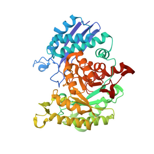
Structure of human alpha-enolase (hENO1), a multifunctional glycolytic enzyme.

Kang, H.J.Jung, S.K.Kim, S.J.Chung, S.J.

(2008) Acta Crystallogr D Biol Crystallogr 64: 651-657

  • DOI: https://doi.org/10.1107/S0907444908008561
  • Primary Citation of Related Structures:  
    3B97

  • PubMed Abstract: 

    Aside from its enzymatic function in the glycolytic pathway, alpha-enolase (ENO1) has been implicated in numerous diseases, including metastatic cancer, autoimmune disorders, ischaemia and bacterial infection. The disease-related roles of ENO1 are mostly attributed to its immunogenic capacity, DNA-binding ability and plasmin(ogen) receptor function, which are significantly affected by its three-dimensional structure and surface properties, rather than its enzymatic activity. Here, the crystal structure of human ENO1 (hENO1) is presented at 2.2 A resolution. Despite its high sequence similarity to other enolases, the hENO1 structure exhibits distinct surface properties, explaining its various activities, including plasmin(ogen) and DNA binding.


  • Organizational Affiliation

    BioNanotechnology Research Center, Korea Research Institute of Bioscience and Biotechnology and Division of Nanobiotechnology, Korea University of Science and Technology (UST), Yuseong, Daejeon 305-333, Republic of Korea.


Macromolecules
Find similar proteins by:  (by identity cutoff)  |  3D Structure
Entity ID: 1
MoleculeChains Sequence LengthOrganismDetailsImage
Alpha-enolase
A, B, C, D
433Homo sapiensMutation(s): 0 
EC: 4.2.1.11
UniProt & NIH Common Fund Data Resources
Find proteins for P06733 (Homo sapiens)
Explore P06733 
Go to UniProtKB:  P06733
PHAROS:  P06733
GTEx:  ENSG00000074800 
Entity Groups  
Sequence Clusters30% Identity50% Identity70% Identity90% Identity95% Identity100% Identity
UniProt GroupP06733
Sequence Annotations
Expand
  • Reference Sequence
Small Molecules
Ligands 2 Unique
IDChains Name / Formula / InChI Key2D Diagram3D Interactions
SO4
Query on SO4

Download Ideal Coordinates CCD File 
G [auth A],
J [auth B],
M [auth C],
P [auth D]
SULFATE ION
O4 S
QAOWNCQODCNURD-UHFFFAOYSA-L
MG
Query on MG

Download Ideal Coordinates CCD File 
E [auth A]
F [auth A]
H [auth B]
I [auth B]
K [auth C]
E [auth A],
F [auth A],
H [auth B],
I [auth B],
K [auth C],
L [auth C],
N [auth D],
O [auth D]
MAGNESIUM ION
Mg
JLVVSXFLKOJNIY-UHFFFAOYSA-N
Experimental Data & Validation

Experimental Data

  • Method: X-RAY DIFFRACTION
  • Resolution: 2.20 Å
  • R-Value Free: 0.217 
  • R-Value Work: 0.184 
  • R-Value Observed: 0.187 
  • Space Group: P 42
Unit Cell:
Length ( Å )Angle ( ˚ )
a = 192.796α = 90
b = 192.796β = 90
c = 65.17γ = 90
Software Package:
Software NamePurpose
HKL-2000data collection
PHASERphasing
CNSrefinement
DENZOdata reduction
SCALEPACKdata scaling

Structure Validation

View Full Validation Report


Entry History 

Deposition Data

Revision History  (Full details and data files)

  • Version 1.0: 2008-09-16
    Type: Initial release
  • Version 1.1: 2011-07-13
    Changes: Version format compliance
  • Version 1.2: 2024-03-13
    Changes: Data collection, Database references, Derived calculations