3AJI

Structure of Gankyrin-S6ATPase photo-cross-linked site-specifically, and incoporated by genetic code expansion


Experimental Data Snapshot

  • Method: X-RAY DIFFRACTION
  • Resolution: 2.05 Å
  • R-Value Free: 0.229 
  • R-Value Work: 0.171 
  • R-Value Observed: 0.171 

wwPDB Validation   3D Report Full Report


This is version 1.4 of the entry. See complete history


Literature

Crystallographic study of a site-specifically cross-linked protein complex with a genetically incorporated photoreactive amino acid

Sato, S.Mimasu, S.Sato, A.Hino, N.Sakamoto, K.Umehara, T.Yokoyama, S.

(2011) Biochemistry 50: 250-257

  • DOI: https://doi.org/10.1021/bi1016183
  • Primary Citation of Related Structures:  
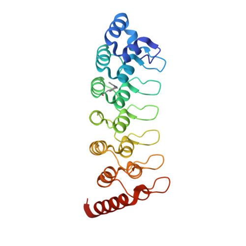
    3AJI

  • PubMed Abstract: 

    The benzophenone photophore is widely used to photo-cross-link macromolecules. Recent developments in genetic code expansion have allowed the biosynthesis of proteins with p-benzoyl-L-phenylalanine (pBpa) at defined sites, for covalent bonding with interacting proteins. However, the structure of a photo-cross-linked protein complex had not been revealed, and thus neither the actual structure of the "photobridge" in a complex nor the influence of this covalent bridge on the overall complex structure was known. In this study, we determine the crystal structure of the cross-linked complex of the liver oncoprotein gankyrin and the C-terminal domain of S6 proteasomal protein (S6C), at 2.05 Å resolution. First, the photoreactive amino acid was separately incorporated into gankyrin at 16 sites on the protein surface, and two variants that efficiently formed a covalent bond with S6C were found. The yield of one of the cross-linked products, with pBpa in place of Arg85 in gankyrin, was maximized for crystallization via optimization of the duration of complex exposure to 365 nm light. The structure revealed that the carbonyl group of the benzophenone of pBpa85 formed a covalent bond exclusively with the Cγ atom of Glu356 in S6C, showing the high selectivity of formation of cross-links by pBpa. In addition, the cross-linked structure exhibited little structural distortion from the native complex structure. Our results demonstrated that cross-linking with site-specifically incorporated pBpa preserves the native binding mode and is useful for probing protein-protein interactions.


  • Organizational Affiliation

    RIKEN Systems and Structural Biology Center, Tsurumi, Yokohama, Japan.


Macromolecules
Find similar proteins by:  (by identity cutoff)  |  3D Structure
Entity ID: 1
MoleculeChains Sequence LengthOrganismDetailsImage
26S proteasome non-ATPase regulatory subunit 10
A, C
231Mus musculusMutation(s): 1 
Gene Names: Psmd10
UniProt
Find proteins for Q9Z2X2 (Mus musculus)
Explore Q9Z2X2 
Go to UniProtKB:  Q9Z2X2
Entity Groups  
Sequence Clusters30% Identity50% Identity70% Identity90% Identity95% Identity100% Identity
UniProt GroupQ9Z2X2
Sequence Annotations
Expand
  • Reference Sequence
Find similar proteins by:  (by identity cutoff)  |  3D Structure
Entity ID: 2
MoleculeChains Sequence LengthOrganismDetailsImage
Proteasome (Prosome, macropain) 26S subunit, ATPase, 4
B, D
83Mus musculusMutation(s): 0 
Gene Names: Psmc4
UniProt
Find proteins for P54775 (Mus musculus)
Explore P54775 
Go to UniProtKB:  P54775
Entity Groups  
Sequence Clusters30% Identity50% Identity70% Identity90% Identity95% Identity100% Identity
UniProt GroupP54775
Sequence Annotations
Expand
  • Reference Sequence
Small Molecules
Modified Residues  1 Unique
IDChains TypeFormula2D DiagramParent
PBF
Query on PBF
A, C
L-PEPTIDE LINKINGC16 H15 N O3PHE
Experimental Data & Validation

Experimental Data

  • Method: X-RAY DIFFRACTION
  • Resolution: 2.05 Å
  • R-Value Free: 0.229 
  • R-Value Work: 0.171 
  • R-Value Observed: 0.171 
  • Space Group: H 3
Unit Cell:
Length ( Å )Angle ( ˚ )
a = 103.164α = 90
b = 103.164β = 90
c = 154.993γ = 120
Software Package:
Software NamePurpose
HKL-2000data collection
MOLREPphasing
CNSrefinement
DENZOdata reduction
SCALEPACKdata scaling

Structure Validation

View Full Validation Report


Entry History 

Revision History  (Full details and data files)

  • Version 1.0: 2010-12-22
    Type: Initial release
  • Version 1.1: 2011-07-13
    Changes: Version format compliance
  • Version 1.2: 2014-01-29
    Changes: Database references
  • Version 1.3: 2023-11-01
    Changes: Data collection, Database references, Derived calculations, Refinement description
  • Version 1.4: 2023-11-15
    Changes: Data collection