3AJ4

Crystal structure of the PH domain of Evectin-2 from human complexed with O-phospho-L-serine


Experimental Data Snapshot

  • Method: X-RAY DIFFRACTION
  • Resolution: 1.00 Å
  • R-Value Free: 0.126 
  • R-Value Work: 0.105 
  • R-Value Observed: 0.106 

wwPDB Validation   3D Report Full Report


Ligand Structure Quality Assessment 


This is version 1.3 of the entry. See complete history


Literature

Intracellular phosphatidylserine is essential for retrograde membrane traffic through endosomes

Uchida, Y.Hasegawa, J.Chinnapen, D.Inoue, T.Okazaki, S.Kato, R.Wakatsuki, S.Misaki, R.Koike, M.Uchiyama, Y.Iemura, S.Natsume, T.Kuwahara, R.Nakagawa, T.Nishikawa, K.Mukai, K.Miyoshi, E.Taniguchi, N.Sheff, D.Lencer, W.I.Taguchi, T.Arai, H.

(2011) Proc Natl Acad Sci U S A 108: 15846-15851

  • DOI: https://doi.org/10.1073/pnas.1109101108
  • Primary Citation of Related Structures:  
    3AJ4

  • PubMed Abstract: 

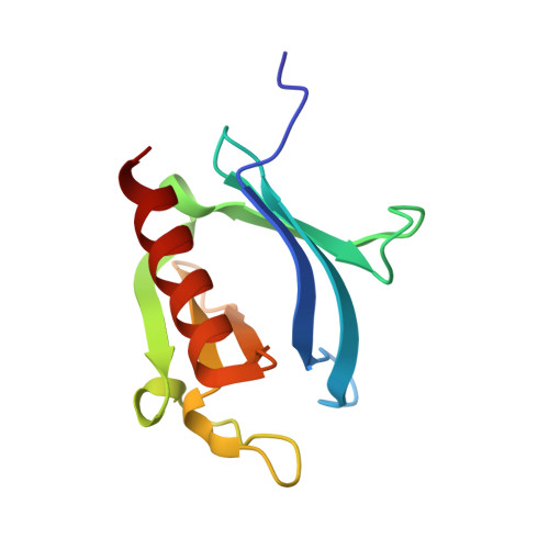
    Phosphatidylserine (PS) is a relatively minor constituent of biological membranes. Despite its low abundance, PS in the plasma membrane (PM) plays key roles in various phenomena such as the coagulation cascade, clearance of apoptotic cells, and recruitment of signaling molecules. PS also localizes in endocytic organelles, but how this relates to its cellular functions remains unknown. Here we report that PS is essential for retrograde membrane traffic at recycling endosomes (REs). PS was most concentrated in REs among intracellular organelles, and evectin-2 (evt-2), a protein of previously unknown function, was targeted to REs by the binding of its pleckstrin homology (PH) domain to PS. X-ray analysis supported the specificity of the binding of PS to the PH domain. Depletion of evt-2 or masking of intracellular PS suppressed membrane traffic from REs to the Golgi. These findings uncover the molecular basis that controls the RE-to-Golgi transport and identify a unique PH domain that specifically recognizes PS but not polyphosphoinositides.


  • Organizational Affiliation

    Department of Health Chemistry, Graduate School of Pharmaceutical Sciences, University of Tokyo, Tokyo 113-0033, Japan.


Macromolecules
Find similar proteins by:  (by identity cutoff)  |  3D Structure
Entity ID: 1
MoleculeChains Sequence LengthOrganismDetailsImage
Pleckstrin homology domain-containing family B member 2
A, B
112Homo sapiensMutation(s): 0 
UniProt & NIH Common Fund Data Resources
Find proteins for Q96CS7 (Homo sapiens)
Explore Q96CS7 
Go to UniProtKB:  Q96CS7
PHAROS:  Q96CS7
GTEx:  ENSG00000115762 
Entity Groups  
Sequence Clusters30% Identity50% Identity70% Identity90% Identity95% Identity100% Identity
UniProt GroupQ96CS7
Sequence Annotations
Expand
  • Reference Sequence
Experimental Data & Validation

Experimental Data

  • Method: X-RAY DIFFRACTION
  • Resolution: 1.00 Å
  • R-Value Free: 0.126 
  • R-Value Work: 0.105 
  • R-Value Observed: 0.106 
  • Space Group: P 1 21 1
Unit Cell:
Length ( Å )Angle ( ˚ )
a = 31.747α = 90
b = 48.449β = 92.2
c = 64.323γ = 90
Software Package:
Software NamePurpose
HKL-2000data collection
MOLREPphasing
REFMACrefinement
HKL-2000data reduction
HKL-2000data scaling

Structure Validation

View Full Validation Report


Ligand Structure Quality Assessment 


Entry History 

Deposition Data

Revision History  (Full details and data files)

  • Version 1.0: 2011-05-25
    Type: Initial release
  • Version 1.1: 2011-07-13
    Changes: Version format compliance
  • Version 1.2: 2011-10-05
    Changes: Database references
  • Version 1.3: 2023-11-01
    Changes: Data collection, Database references, Derived calculations, Refinement description