3AEQ

Structure of the light-independent protochlorophyllide reductase catalyzing a key reduction for greening in the dark


Experimental Data Snapshot

  • Method: X-RAY DIFFRACTION
  • Resolution: 2.90 Å
  • R-Value Free: 0.298 
  • R-Value Work: 0.235 
  • R-Value Observed: 0.238 

wwPDB Validation   3D Report Full Report


Ligand Structure Quality Assessment 


This is version 1.2 of the entry. See complete history


Literature

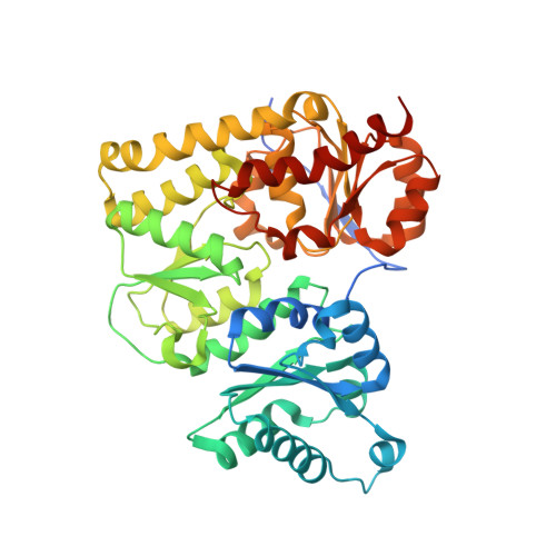
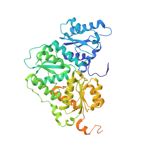
X-ray crystal structure of the light-independent protochlorophyllide reductase

Muraki, N.Nomata, J.Ebata, K.Mizoguchi, T.Shiba, T.Tamiaki, H.Kurisu, G.Fujita, Y.

(2010) Nature 465: 110-114

  • DOI: https://doi.org/10.1038/nature08950
  • Primary Citation of Related Structures:  
    3AEK, 3AEQ, 3AER, 3AES, 3AET, 3AEU

  • PubMed Abstract: 

    Photosynthetic organisms adopt two different strategies for the reduction of the C17 = C18 double bond of protochlorophyllide (Pchlide) to form chlorophyllide a, the direct precursor of chlorophyll a (refs 1-4). The first involves the activity of the light-dependent Pchlide oxidoreductase, and the second involves the light-independent (dark-operative) Pchlide oxidoreductase (DPOR). DPOR is a nitrogenase-like enzyme consisting of two components, L-protein (a BchL dimer) and NB-protein (a BchN-BchB heterotetramer), which are structurally related to nitrogenase Fe protein and MoFe protein, respectively. Here we report the crystal structure of the NB-protein of DPOR from Rhodobacter capsulatus at a resolution of 2.3A. As expected, the overall structure is similar to that of nitrogenase MoFe protein: each catalytic BchN-BchB unit contains one Pchlide and one iron-sulphur cluster (NB-cluster) coordinated uniquely by one aspartate and three cysteines. Unique aspartate ligation is not necessarily needed for the cluster assembly but is essential for the catalytic activity. Specific Pchlide-binding accompanies the partial unwinding of an alpha-helix that belongs to the next catalytic BchN-BchB unit. We propose a unique trans-specific reduction mechanism in which the distorted C17-propionate of Pchlide and an aspartate from BchB serve as proton donors for C18 and C17 of Pchlide, respectively. Intriguingly, the spatial arrangement of the NB-cluster and Pchlide is almost identical to that of the P-cluster and FeMo-cofactor in nitrogenase MoFe-protein, illustrating that a common architecture exists to reduce chemically stable multibonds of porphyrin and dinitrogen.


  • Organizational Affiliation

    Department of Life Sciences, University of Tokyo, Komaba, Meguro-ku, Tokyo 153-8902, Japan.


Macromolecules
Find similar proteins by:  (by identity cutoff)  |  3D Structure
Entity ID: 1
MoleculeChains Sequence LengthOrganismDetailsImage
Light-independent protochlorophyllide reductase subunit N
A, C
437Rhodobacter capsulatusMutation(s): 0 
Gene Names: bchN
EC: 1.18
UniProt
Find proteins for P26164 (Rhodobacter capsulatus (strain ATCC BAA-309 / NBRC 16581 / SB1003))
Explore P26164 
Go to UniProtKB:  P26164
Entity Groups  
Sequence Clusters30% Identity50% Identity70% Identity90% Identity95% Identity100% Identity
UniProt GroupP26164
Sequence Annotations
Expand
  • Reference Sequence
Find similar proteins by:  (by identity cutoff)  |  3D Structure
Entity ID: 2
MoleculeChains Sequence LengthOrganismDetailsImage
Light-independent protochlorophyllide reductase subunit B
B, D
525Rhodobacter capsulatusMutation(s): 0 
Gene Names: bchBbchK
EC: 1.18
UniProt
Find proteins for P26163 (Rhodobacter capsulatus (strain ATCC BAA-309 / NBRC 16581 / SB1003))
Explore P26163 
Go to UniProtKB:  P26163
Entity Groups  
Sequence Clusters30% Identity50% Identity70% Identity90% Identity95% Identity100% Identity
UniProt GroupP26163
Sequence Annotations
Expand
  • Reference Sequence
Experimental Data & Validation

Experimental Data

  • Method: X-RAY DIFFRACTION
  • Resolution: 2.90 Å
  • R-Value Free: 0.298 
  • R-Value Work: 0.235 
  • R-Value Observed: 0.238 
  • Space Group: P 1 21 1
Unit Cell:
Length ( Å )Angle ( ˚ )
a = 81.632α = 90
b = 81.278β = 100.43
c = 177.103γ = 90
Software Package:
Software NamePurpose
MOLREPphasing
REFMACrefinement
HKL-2000data reduction
HKL-2000data scaling

Structure Validation

View Full Validation Report


Ligand Structure Quality Assessment 


Entry History 

Deposition Data

Revision History  (Full details and data files)

  • Version 1.0: 2010-04-21
    Type: Initial release
  • Version 1.1: 2011-07-13
    Changes: Advisory, Version format compliance
  • Version 1.2: 2023-11-01
    Changes: Data collection, Database references, Derived calculations, Refinement description