3ZUX

Crystal structure of a bacterial homologue of the bile acid sodium symporter ASBT.


Experimental Data Snapshot

  • Method: X-RAY DIFFRACTION
  • Resolution: 2.20 Å
  • R-Value Free: 0.229 
  • R-Value Work: 0.197 
  • R-Value Observed: 0.199 

wwPDB Validation   3D Report Full Report


Ligand Structure Quality Assessment 


This is version 1.2 of the entry. See complete history


Literature

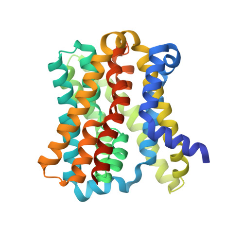
Crystal Structure of a Bacterial Homologue of the Bile Acid Sodium Symporter Asbt.

Hu, N.-J.Iwata, S.Cameron, A.D.Drew, D.

(2011) Nature 478: 408

  • DOI: https://doi.org/10.1038/nature10450
  • Primary Citation of Related Structures:  
    3ZUX, 3ZUY

  • PubMed Abstract: 

    High cholesterol levels greatly increase the risk of cardiovascular disease. About 50 per cent of cholesterol is eliminated from the body by its conversion into bile acids. However, bile acids released from the bile duct are constantly recycled, being reabsorbed in the intestine by the apical sodium-dependent bile acid transporter (ASBT, also known as SLC10A2). It has been shown in animal models that plasma cholesterol levels are considerably lowered by specific inhibitors of ASBT, and ASBT is thus a target for hypercholesterolaemia drugs. Here we report the crystal structure of a bacterial homologue of ASBT from Neisseria meningitidis (ASBT(NM)) at 2.2 Å. ASBT(NM) contains two inverted structural repeats of five transmembrane helices. A core domain of six helices harbours two sodium ions, and the remaining four helices pack in a row to form a flat, 'panel'-like domain. Overall, the architecture of the protein is remarkably similar to the sodium/proton antiporter NhaA, despite having no detectable sequence homology. The ASBT(NM) structure was captured with the substrate taurocholate present, bound between the core and panel domains in a large, inward-facing, hydrophobic cavity. Residues near this cavity have been shown to affect the binding of specific inhibitors of human ASBT. The position of the taurocholate molecule, together with the molecular architecture, suggests the rudiments of a possible transport mechanism.


  • Organizational Affiliation

    Division of Molecular Biosciences, Imperial College London, London SW7 2AZ, UK.


Macromolecules
Find similar proteins by:  (by identity cutoff)  |  3D Structure
Entity ID: 1
MoleculeChains Sequence LengthOrganismDetailsImage
Transporter332Neisseria meningitidisMutation(s): 3 
Gene Names: NMB0705
Membrane Entity: Yes 
UniProt
Find proteins for Q9K0A9 (Neisseria meningitidis serogroup B (strain MC58))
Explore Q9K0A9 
Go to UniProtKB:  Q9K0A9
Entity Groups  
Sequence Clusters30% Identity50% Identity70% Identity90% Identity95% Identity100% Identity
UniProt GroupQ9K0A9
Sequence Annotations
Expand
  • Reference Sequence
Small Molecules
Ligands 5 Unique
IDChains Name / Formula / InChI Key2D Diagram3D Interactions
PTY
Query on PTY

Download Ideal Coordinates CCD File 
J [auth A],
K [auth A]
PHOSPHATIDYLETHANOLAMINE
C40 H80 N O8 P
NJGIRBISCGPRPF-KXQOOQHDSA-N
TCH
Query on TCH

Download Ideal Coordinates CCD File 
E [auth A],
F [auth A]
TAUROCHOLIC ACID
C26 H45 N O7 S
WBWWGRHZICKQGZ-HZAMXZRMSA-N
LDA
Query on LDA

Download Ideal Coordinates CCD File 
G [auth A],
H [auth A],
I [auth A],
L [auth A],
M [auth A]
LAURYL DIMETHYLAMINE-N-OXIDE
C14 H31 N O
SYELZBGXAIXKHU-UHFFFAOYSA-N
HG
Query on HG

Download Ideal Coordinates CCD File 
D [auth A]MERCURY (II) ION
Hg
BQPIGGFYSBELGY-UHFFFAOYSA-N
NA
Query on NA

Download Ideal Coordinates CCD File 
B [auth A],
C [auth A]
SODIUM ION
Na
FKNQFGJONOIPTF-UHFFFAOYSA-N
Experimental Data & Validation

Experimental Data

  • Method: X-RAY DIFFRACTION
  • Resolution: 2.20 Å
  • R-Value Free: 0.229 
  • R-Value Work: 0.197 
  • R-Value Observed: 0.199 
  • Space Group: P 41 2 2
Unit Cell:
Length ( Å )Angle ( ˚ )
a = 73.33α = 90
b = 73.33β = 90
c = 163.52γ = 90
Software Package:
Software NamePurpose
BUSTERrefinement
MOSFLMdata reduction
SCALAdata scaling
SHARPphasing

Structure Validation

View Full Validation Report


Ligand Structure Quality Assessment 


Entry History 

Deposition Data

Revision History  (Full details and data files)

  • Version 1.0: 2011-10-12
    Type: Initial release
  • Version 1.1: 2011-10-26
    Changes: Database references, Structure summary
  • Version 1.2: 2023-05-31
    Changes: Database references, Derived calculations, Other, Refinement description, Source and taxonomy, Structure summary