3ZL8

CRYSTAL STRUCTURE OF MURF LIGASE FROM THERMOTOGA MARITIMA IN COMPLEX WITH ADP


Experimental Data Snapshot

  • Method: X-RAY DIFFRACTION
  • Resolution: 1.65 Å
  • R-Value Free: 0.220 
  • R-Value Work: 0.164 
  • R-Value Observed: 0.169 

wwPDB Validation   3D Report Full Report


Ligand Structure Quality Assessment 


This is version 1.1 of the entry. See complete history


Literature

Mreb and Murg as Scaffolds for the Cytoplasmic Steps of Peptidoglycan Biosynthesis

Favini-Stabile, S.Contreras-Martel, C.Thielens, N.Dessen, A.

(2013) Environ Microbiol 15: 3218

  • DOI: https://doi.org/10.1111/1462-2920.12171
  • Primary Citation of Related Structures:  
    3ZL8, 4BUB, 4BUC

  • PubMed Abstract: 

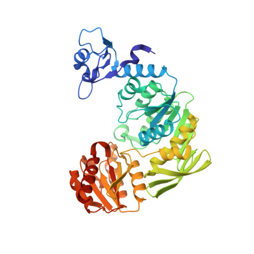
    Peptidoglycan is a major determinant of cell shape in bacteria, and its biosynthesis involves the concerted action of cytoplasmic, membrane-associated and periplasmic enzymes. Within the cytoplasm, Mur enzymes catalyse the first steps leading to peptidoglycan precursor biosynthesis, and have been suggested as being part of a multicomponent complex that could also involve the transglycosylase MurG and the cytoskeletal protein MreB. In order to initialize the characterization of a potential Mur interaction network, we purified MurD, MurE, MurF, MurG and MreB from Thermotoga maritima and characterized their interactions using membrane blotting and surface plasmon resonance. MurD, MurE and MurF all recognize MurG and MreB, but not each other, while the two latter proteins interact. In addition, we solved the crystal structures of MurD, MurE and MurF, which indicate that their C-termini display high conformational flexibilities. The differences in Mur conformations could be important parameters for the stability of an intracytoplasmic murein biosynthesis complex.


  • Organizational Affiliation

    Institut de Biologie Structurale (IBS), Université Grenoble I, Grenoble, France; Commissariat à l'Energie Atomique (CEA), Grenoble, France; Centre National de la Recherche Scientifique (CNRS), Grenoble, France.


Macromolecules
Find similar proteins by:  (by identity cutoff)  |  3D Structure
Entity ID: 1
MoleculeChains Sequence LengthOrganismDetailsImage
UDP-N-ACETYLMURAMOYL-TRIPEPTIDE--D-ALANYL-D-ALANINE LIGASE437Thermotoga maritimaMutation(s): 0 
EC: 6.3.2.10
UniProt
Find proteins for Q9WY78 (Thermotoga maritima (strain ATCC 43589 / DSM 3109 / JCM 10099 / NBRC 100826 / MSB8))
Explore Q9WY78 
Go to UniProtKB:  Q9WY78
Entity Groups  
Sequence Clusters30% Identity50% Identity70% Identity90% Identity95% Identity100% Identity
UniProt GroupQ9WY78
Sequence Annotations
Expand
  • Reference Sequence
Small Molecules
Ligands 2 Unique
IDChains Name / Formula / InChI Key2D Diagram3D Interactions
ADP
Query on ADP

Download Ideal Coordinates CCD File 
C [auth A]ADENOSINE-5'-DIPHOSPHATE
C10 H15 N5 O10 P2
XTWYTFMLZFPYCI-KQYNXXCUSA-N
GOL
Query on GOL

Download Ideal Coordinates CCD File 
B [auth A]GLYCEROL
C3 H8 O3
PEDCQBHIVMGVHV-UHFFFAOYSA-N
Experimental Data & Validation

Experimental Data

  • Method: X-RAY DIFFRACTION
  • Resolution: 1.65 Å
  • R-Value Free: 0.220 
  • R-Value Work: 0.164 
  • R-Value Observed: 0.169 
  • Space Group: P 21 21 21
Unit Cell:
Length ( Å )Angle ( ˚ )
a = 48.229α = 90
b = 49.595β = 90
c = 177.164γ = 90
Software Package:
Software NamePurpose
REFMACrefinement
XDSdata reduction
XSCALEdata scaling
autoSHARPphasing

Structure Validation

View Full Validation Report


Ligand Structure Quality Assessment 


Entry History 

Deposition Data

Revision History  (Full details and data files)

  • Version 1.0: 2013-09-11
    Type: Initial release
  • Version 1.1: 2013-12-18
    Changes: Database references