3ZD5

THE 2.2 A STRUCTURE OF A FULL-LENGTH CATALYTICALLY ACTIVE HAMMERHEAD RIBOZYME


Experimental Data Snapshot

  • Method: X-RAY DIFFRACTION
  • Resolution: 2.20 Å
  • R-Value Free: 0.212 
  • R-Value Work: 0.177 
  • R-Value Observed: 0.180 

wwPDB Validation   3D Report Full Report


This is version 1.2 of the entry. See complete history


Literature

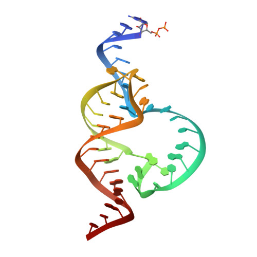
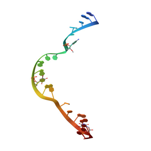
Tertiary contacts distant from the active site prime a ribozyme for catalysis.

Martick, M.Scott, W.G.

(2006) Cell 126: 309-320

  • DOI: https://doi.org/10.1016/j.cell.2006.06.036
  • Primary Citation of Related Structures:  
    3ZD5

  • PubMed Abstract: 

    Minimal hammerhead ribozymes have been characterized extensively by static and time-resolved crystallography as well as numerous biochemical analyses, leading to mutually contradictory mechanistic explanations for catalysis. We present the 2.2 A resolution crystal structure of a full-length Schistosoma mansoni hammerhead ribozyme that permits us to explain the structural basis for its 1000-fold catalytic enhancement. The full-length hammerhead structure reveals how tertiary interactions occurring remotely from the active site prime this ribozyme for catalysis. G-12 and G-8 are positioned consistent with their previously suggested roles in acid-base catalysis, the nucleophile is aligned with a scissile phosphate positioned proximal to the A-9 phosphate, and previously unexplained roles of other conserved nucleotides become apparent within the context of a distinctly new fold that nonetheless accommodates the previous structural studies. These interactions permit us to explain the previously irreconcilable sets of experimental results in a unified, consistent, and unambiguous manner.


  • Organizational Affiliation

    Department of Molecular, Cellular and Developmental Biology, Robert L. Sinsheimer Laboratories, University of California, Santa Cruz, Santa Cruz, CA 95064, USA.


Macromolecules
Find similar nucleic acids by:  (by identity cutoff)  |  3D Structure
Entity ID: 1
MoleculeChains LengthOrganismImage
HAMMERHEAD RIBOZYME, ENZYME STRAND43Schistosoma mansoni
Sequence Annotations
Expand
  • Reference Sequence

Find similar nucleic acids by:  Sequence   |   3D Structure  

Entity ID: 2
MoleculeChains LengthOrganismImage
HAMMERHEAD RIBOZYME, SUBSTRATE STRAND20Schistosoma mansoni
Sequence Annotations
Expand
  • Reference Sequence
Experimental Data & Validation

Experimental Data

  • Method: X-RAY DIFFRACTION
  • Resolution: 2.20 Å
  • R-Value Free: 0.212 
  • R-Value Work: 0.177 
  • R-Value Observed: 0.180 
  • Space Group: C 1 2 1
Unit Cell:
Length ( Å )Angle ( ˚ )
a = 49.886α = 90
b = 69.21β = 111.83
c = 60.076γ = 90
Software Package:
Software NamePurpose
PHENIXrefinement
ADSCdata reduction
MOSFLMdata scaling
CNSphasing

Structure Validation

View Full Validation Report


Entry History 

Deposition Data

Revision History  (Full details and data files)

  • Version 1.0: 2012-12-12
    Type: Initial release
  • Version 1.1: 2013-03-06
    Changes: Other
  • Version 1.2: 2019-03-13
    Changes: Data collection, Database references, Derived calculations, Experimental preparation, Other, Source and taxonomy