3WYQ

Crystal structure of the low-immunogenic core streptavidin mutant LISA-314 (Y22S/Y83S/R84K/E101D/R103K/E116N) at 1.0 A resolution


Experimental Data Snapshot

  • Method: X-RAY DIFFRACTION
  • Resolution: 1.00 Å
  • R-Value Free: 0.163 
  • R-Value Work: 0.137 
  • R-Value Observed: 0.138 

wwPDB Validation   3D Report Full Report


Ligand Structure Quality Assessment 


This is version 1.1 of the entry. See complete history


Literature

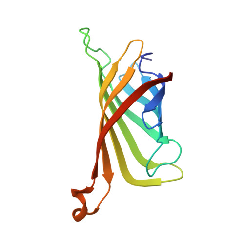
Crystal structure of streptavidin mutant with low immunogenicity.

Kawato, T.Mizohata, E.Meshizuka, T.Doi, H.Kawamura, T.Matsumura, H.Yumura, K.Tsumoto, K.Kodama, T.Inoue, T.Sugiyama, A.

(2015) J Biosci Bioeng 119: 642-647

  • DOI: https://doi.org/10.1016/j.jbiosc.2014.10.025
  • Primary Citation of Related Structures:  
    3WYP, 3WYQ

  • PubMed Abstract: 

    We previously created a low-immunogenic core streptavidin mutant No. 314 (LISA-314) by replacing six amino-acid residues for use as a delivery tool for an antibody multistep pre-targeting process (Yumura et al., Protein Sci., 22, 213-221, 2013). Here, we performed high-resolution X-ray structural analyses of LISA-314 and wild-type streptavidin to investigate the effect of substitutions on the protein function and the three-dimensional structure. LISA-314 forms a tetramer in the same manner as wild-type streptavidin. The binding mode of d-biotin in LISA-314 is also completely identical to that in wild-type streptavidin, and conformational changes were observed mostly at the side chains of substituted sites. Any large conformational changes corresponding to the reduction of B factors around the substituted sites were not observed. These results demonstrated the LISA-314 acquired low immunogenicity without losing structural properties of original wild-type streptavidin.


  • Organizational Affiliation

    Division of Applied Chemistry, Graduate School of Engineering, Osaka University, 2-1 Yamadaoka, Suita, Osaka 565-0871, Japan.


Macromolecules
Find similar proteins by:  (by identity cutoff)  |  3D Structure
Entity ID: 1
MoleculeChains Sequence LengthOrganismDetailsImage
Streptavidin
A, B
127Streptomyces avidiniiMutation(s): 6 
UniProt
Find proteins for P22629 (Streptomyces avidinii)
Explore P22629 
Go to UniProtKB:  P22629
Entity Groups  
Sequence Clusters30% Identity50% Identity70% Identity90% Identity95% Identity100% Identity
UniProt GroupP22629
Sequence Annotations
Expand
  • Reference Sequence
Small Molecules
Ligands 3 Unique
IDChains Name / Formula / InChI Key2D Diagram3D Interactions
BTN
Query on BTN

Download Ideal Coordinates CCD File 
C [auth A],
K [auth B]
BIOTIN
C10 H16 N2 O3 S
YBJHBAHKTGYVGT-ZKWXMUAHSA-N
SO4
Query on SO4

Download Ideal Coordinates CCD File 
J [auth A]SULFATE ION
O4 S
QAOWNCQODCNURD-UHFFFAOYSA-L
GOL
Query on GOL

Download Ideal Coordinates CCD File 
D [auth A]
E [auth A]
F [auth A]
G [auth A]
H [auth A]
D [auth A],
E [auth A],
F [auth A],
G [auth A],
H [auth A],
I [auth A],
L [auth B],
M [auth B],
N [auth B],
O [auth B]
GLYCEROL
C3 H8 O3
PEDCQBHIVMGVHV-UHFFFAOYSA-N
Binding Affinity Annotations 
IDSourceBinding Affinity
BTN BindingDB:  3WYQ Kd: 1 (nM) from 1 assay(s)
ΔH: min: -1.23e+2, max: -6.69e+1 (kJ/mol) from 12 assay(s)
Experimental Data & Validation

Experimental Data

  • Method: X-RAY DIFFRACTION
  • Resolution: 1.00 Å
  • R-Value Free: 0.163 
  • R-Value Work: 0.137 
  • R-Value Observed: 0.138 
  • Space Group: C 1 2 1
Unit Cell:
Length ( Å )Angle ( ˚ )
a = 63.223α = 90
b = 67.066β = 116.68
c = 56.363γ = 90
Software Package:
Software NamePurpose
BSSdata collection
PHASERphasing
REFMACrefinement
HKL-2000data reduction
HKL-2000data scaling

Structure Validation

View Full Validation Report


Ligand Structure Quality Assessment 


Entry History 

Deposition Data

Revision History  (Full details and data files)

  • Version 1.0: 2014-12-24
    Type: Initial release
  • Version 1.1: 2022-08-24
    Changes: Database references, Derived calculations