3WSO

Crystal structure of the Skp1-FBG3 complex


Experimental Data Snapshot

  • Method: X-RAY DIFFRACTION
  • Resolution: 2.60 Å
  • R-Value Free: 0.266 
  • R-Value Work: 0.205 
  • R-Value Observed: 0.208 

wwPDB Validation   3D Report Full Report


This is version 1.3 of the entry. See complete history


Literature

The Structural Differences between a Glycoprotein Specific F-Box Protein Fbs1 and Its Homologous Protein FBG3

Kumanomidou, T.Nishio, K.Takagi, K.Nakagawa, T.Suzuki, A.Yamane, T.Tokunaga, F.Iwai, K.Murakami, A.Yoshida, Y.Tanaka, K.Mizushima, T.

(2015) PLoS One 10: e0140366-e0140366

  • DOI: https://doi.org/10.1371/journal.pone.0140366
  • Primary Citation of Related Structures:  
    3WSO

  • PubMed Abstract: 

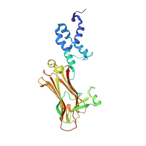
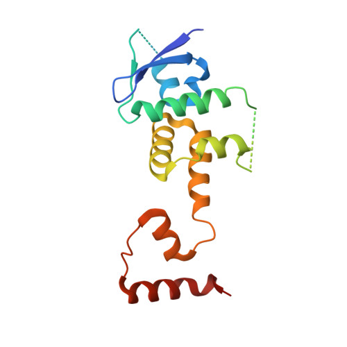
    The Skp1-Cul1-F-box protein (SCF) complex catalyzes protein ubiquitination in diverse cellular processes and is one of the best-characterized ubiquitin ligases. F-box proteins determine the substrate specificities of SCF ubiquitin ligases. Among these, Fbs1/FBG1/FBXO2, Fbs2/FBG2/FBXO6, and Fbs3/FBG5/FBXO27 recognize the N-glycans of glycoproteins, whereas FBG3/FBXO44 has no sugar-binding activity, despite the high sequence homology and conservation of the residues necessary for oligosaccharide binding between Fbs1-3 and FBG3. Here we determined the crystal structure of the Skp1-FBG3 complex at a resolution of 2.6 Å. The substrate-binding domain of FBG3 is composed of a 10-stranded antiparallel β-sandwich with three helices. Although the overall structure of FBG3 is similar to that of Fbs1, the residues that form the Fbs1 carbohydrate-binding pocket failed to be superposed with the corresponding residues of FBG3. Structure-based mutational analysis shows that distinct hydrogen bond networks of four FBG3 loops, i.e., β2-β3, β5-β6, β7-β8, and β9-β10, prevent the formation of the carbohydrate-binding pocket shown in Fbs1.


  • Organizational Affiliation

    Department of Biotechnology, Graduate School of Engineering, Nagoya University, Chikusa-ku, Nagoya, Japan.


Macromolecules
Find similar proteins by:  (by identity cutoff)  |  3D Structure
Entity ID: 1
MoleculeChains Sequence LengthOrganismDetailsImage
F-box only protein 44255Homo sapiensMutation(s): 0 
Gene Names: FBXO44FBG3FBX30FBX44FBX6AFBXO6A
UniProt & NIH Common Fund Data Resources
Find proteins for Q9H4M3 (Homo sapiens)
Explore Q9H4M3 
Go to UniProtKB:  Q9H4M3
PHAROS:  Q9H4M3
GTEx:  ENSG00000132879 
Entity Groups  
Sequence Clusters30% Identity50% Identity70% Identity90% Identity95% Identity100% Identity
UniProt GroupQ9H4M3
Sequence Annotations
Expand
  • Reference Sequence
Find similar proteins by:  (by identity cutoff)  |  3D Structure
Entity ID: 2
MoleculeChains Sequence LengthOrganismDetailsImage
S-phase kinase-associated protein 1166Homo sapiensMutation(s): 0 
Gene Names: SKP1EMC19OCP2SKP1ATCEB1L
UniProt & NIH Common Fund Data Resources
Find proteins for P63208 (Homo sapiens)
Explore P63208 
Go to UniProtKB:  P63208
PHAROS:  P63208
GTEx:  ENSG00000113558 
Entity Groups  
Sequence Clusters30% Identity50% Identity70% Identity90% Identity95% Identity100% Identity
UniProt GroupP63208
Sequence Annotations
Expand
  • Reference Sequence
Experimental Data & Validation

Experimental Data

  • Method: X-RAY DIFFRACTION
  • Resolution: 2.60 Å
  • R-Value Free: 0.266 
  • R-Value Work: 0.205 
  • R-Value Observed: 0.208 
  • Space Group: P 21 21 21
Unit Cell:
Length ( Å )Angle ( ˚ )
a = 34.12α = 90
b = 76.594β = 90
c = 193.939γ = 90
Software Package:
Software NamePurpose
MOLREPphasing
REFMACrefinement
DENZOdata reduction
SCALEPACKdata scaling

Structure Validation

View Full Validation Report


Entry History 

Revision History  (Full details and data files)

  • Version 1.0: 2015-03-25
    Type: Initial release
  • Version 1.1: 2015-10-28
    Changes: Database references
  • Version 1.2: 2015-11-11
    Changes: Other
  • Version 1.3: 2023-11-08
    Changes: Data collection, Database references, Refinement description