3WSJ

HTLV-1 protease in complex with the HIV-1 protease inhibitor Indinavir


Experimental Data Snapshot

  • Method: X-RAY DIFFRACTION
  • Resolution: 2.40 Å
  • R-Value Free: 0.256 
  • R-Value Work: 0.205 
  • R-Value Observed: 0.209 

wwPDB Validation   3D Report Full Report


Ligand Structure Quality Assessment 


This is version 1.1 of the entry. See complete history


Literature

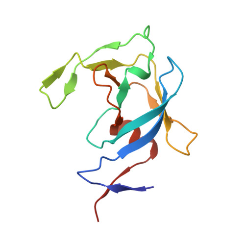
Structural basis for HTLV-1 protease inhibition by the HIV-1 protease inhibitor indinavir.

Kuhnert, M.Steuber, H.Diederich, W.E.

(2014) J Med Chem 57: 6266-6272

  • DOI: https://doi.org/10.1021/jm500402c
  • Primary Citation of Related Structures:  
    3WSJ

  • PubMed Abstract: 

    HTLV-1 protease (HTLV-1 PR) is an aspartic protease which represents a promising drug target for the discovery of novel anti-HTLV-1 drugs. The X-ray structure of HTLV-1 PR in complex with the well-known and approved HIV-1 PR inhibitor Indinavir was determined at 2.40 Å resolution. In this contribution, we describe the first crystal structure in complex with a nonpeptidic inhibitor that accounts for rationalizing the rather moderate affinity of Indinavir against HTLV-1 PR and provides the basis for further structure-guided optimization strategies.


  • Organizational Affiliation

    Institut für Pharmazeutische Chemie, Philipps-Universität Marburg , Marbacher Weg 6, 35032 Marburg, Germany.


Macromolecules
Find similar proteins by:  (by identity cutoff)  |  3D Structure
Entity ID: 1
MoleculeChains Sequence LengthOrganismDetailsImage
Protease
A, B
116Human T-cell leukemia virus type IMutation(s): 1 
Gene Names: prt
UniProt
Find proteins for Q82134 (Human T-cell leukemia virus type I)
Explore Q82134 
Go to UniProtKB:  Q82134
Entity Groups  
Sequence Clusters30% Identity50% Identity70% Identity90% Identity95% Identity100% Identity
UniProt GroupQ82134
Sequence Annotations
Expand
  • Reference Sequence
Small Molecules
Ligands 3 Unique
IDChains Name / Formula / InChI Key2D Diagram3D Interactions
MK1
Query on MK1

Download Ideal Coordinates CCD File 
C [auth A]N-[2(R)-HYDROXY-1(S)-INDANYL]-5-[(2(S)-TERTIARY BUTYLAMINOCARBONYL)-4(3-PYRIDYLMETHYL)PIPERAZINO]-4(S)-HYDROXY-2(R)-PHENYLMETHYLPENTANAMIDE
C36 H47 N5 O4
CBVCZFGXHXORBI-PXQQMZJSSA-N
PG4
Query on PG4

Download Ideal Coordinates CCD File 
N [auth A]TETRAETHYLENE GLYCOL
C8 H18 O5
UWHCKJMYHZGTIT-UHFFFAOYSA-N
SO4
Query on SO4

Download Ideal Coordinates CCD File 
D [auth A]
E [auth A]
F [auth A]
G [auth A]
H [auth A]
D [auth A],
E [auth A],
F [auth A],
G [auth A],
H [auth A],
I [auth A],
J [auth A],
K [auth A],
L [auth A],
M [auth A],
O [auth B],
P [auth B],
Q [auth B]
SULFATE ION
O4 S
QAOWNCQODCNURD-UHFFFAOYSA-L
Binding Affinity Annotations 
IDSourceBinding Affinity
MK1 Binding MOAD:  3WSJ Ki: 3.50e+4 (nM) from 1 assay(s)
Experimental Data & Validation

Experimental Data

  • Method: X-RAY DIFFRACTION
  • Resolution: 2.40 Å
  • R-Value Free: 0.256 
  • R-Value Work: 0.205 
  • R-Value Observed: 0.209 
  • Space Group: P 63 2 2
Unit Cell:
Length ( Å )Angle ( ˚ )
a = 77.26α = 90
b = 77.26β = 90
c = 159.34γ = 120
Software Package:
Software NamePurpose
MxCuBEdata collection
PHASERphasing
PHENIXrefinement
XDSdata reduction
XSCALEdata scaling

Structure Validation

View Full Validation Report


Ligand Structure Quality Assessment 


Entry History 

Deposition Data

Revision History  (Full details and data files)

  • Version 1.0: 2014-10-22
    Type: Initial release
  • Version 1.1: 2023-11-08
    Changes: Data collection, Database references, Derived calculations, Refinement description