3WN2

Crystal Structure of Streptomyces coelicolor alpha-L-arabinofuranosidase in complex with xylohexaose


Experimental Data Snapshot

  • Method: X-RAY DIFFRACTION
  • Resolution: 2.10 Å
  • R-Value Free: 0.191 
  • R-Value Work: 0.167 
  • R-Value Observed: 0.168 

wwPDB Validation   3D Report Full Report


This is version 2.1 of the entry. See complete history


Literature

Crystal structure and characterization of the glycoside hydrolase family 62 alpha-L-arabinofuranosidase from Streptomyces coelicolor

Maehara, T.Fujimoto, Z.Ichinose, H.Michikawa, M.Harazono, K.Kaneko, S.

(2014) J Biol Chem 289: 7962-7972

  • DOI: https://doi.org/10.1074/jbc.M113.540542
  • Primary Citation of Related Structures:  
    3WMY, 3WMZ, 3WN0, 3WN1, 3WN2

  • PubMed Abstract: 

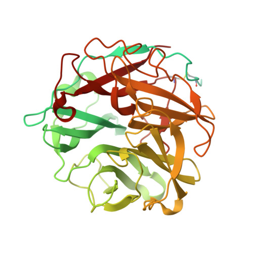
    α-L-arabinofuranosidase, which belongs to the glycoside hydrolase family 62 (GH62), hydrolyzes arabinoxylan but not arabinan or arabinogalactan. The crystal structures of several α-L-arabinofuranosidases have been determined, although the structures, catalytic mechanisms, and substrate specificities of GH62 enzymes remain unclear. To evaluate the substrate specificity of a GH62 enzyme, we determined the crystal structure of α-L-arabinofuranosidase, which comprises a carbohydrate-binding module family 13 domain at its N terminus and a catalytic domain at its C terminus, from Streptomyces coelicolor. The catalytic domain was a five-bladed β-propeller consisting of five radially oriented anti-parallel β-sheets. Sugar complex structures with l-arabinose, xylotriose, and xylohexaose revealed five subsites in the catalytic cleft and an l-arabinose-binding pocket at the bottom of the cleft. The entire structure of this GH62 family enzyme was very similar to that of glycoside hydrolase 43 family enzymes, and the catalytically important acidic residues found in family 43 enzymes were conserved in GH62. Mutagenesis studies revealed that Asp(202) and Glu(361) were catalytic residues, and Trp(270), Tyr(461), and Asn(462) were involved in the substrate-binding site for discriminating the substrate structures. In particular, hydrogen bonding between Asn(462) and xylose at the nonreducing end subsite +2 was important for the higher activity of substituted arabinofuranosyl residues than that for terminal arabinofuranoses.


  • Organizational Affiliation

    From the Food Biotechnology Division, National Food Research Institute, 2-1-12 Kannondai, Tsukuba, Ibaraki 305-8642.


Macromolecules
Find similar proteins by:  (by identity cutoff)  |  3D Structure
Entity ID: 1
MoleculeChains Sequence LengthOrganismDetailsImage
Extracellular exo-alpha-L-arabinofuranosidase438Streptomyces coelicolor A3(2)Mutation(s): 0 
Gene Names: abfBSC7H1.02SCO5932
EC: 3.2.1.55
UniProt
Find proteins for O54161 (Streptomyces coelicolor (strain ATCC BAA-471 / A3(2) / M145))
Explore O54161 
Go to UniProtKB:  O54161
Entity Groups  
Sequence Clusters30% Identity50% Identity70% Identity90% Identity95% Identity100% Identity
UniProt GroupO54161
Sequence Annotations
Expand
  • Reference Sequence
Oligosaccharides

Help

Entity ID: 2
MoleculeChains Length2D Diagram Glycosylation3D Interactions
beta-D-xylopyranose-(1-4)-beta-D-xylopyranose-(1-4)-beta-D-xylopyranose-(1-4)-beta-D-xylopyranose-(1-4)-beta-D-xylopyranose
B
5N/A
Glycosylation Resources
GlyTouCan:  G47101GE
GlyCosmos:  G47101GE
Experimental Data & Validation

Experimental Data

  • Method: X-RAY DIFFRACTION
  • Resolution: 2.10 Å
  • R-Value Free: 0.191 
  • R-Value Work: 0.167 
  • R-Value Observed: 0.168 
  • Space Group: P 41 21 2
Unit Cell:
Length ( Å )Angle ( ˚ )
a = 97.437α = 90
b = 97.437β = 90
c = 103.242γ = 90
Software Package:
Software NamePurpose
SERGUIdata collection
REFMACrefinement
HKL-2000data reduction
SCALEPACKdata scaling
REFMACphasing

Structure Validation

View Full Validation Report


Entry History 

Deposition Data

Revision History  (Full details and data files)

  • Version 1.0: 2014-02-05
    Type: Initial release
  • Version 1.1: 2014-10-29
    Changes: Database references
  • Version 1.2: 2017-11-22
    Changes: Refinement description
  • Version 2.0: 2020-07-29
    Type: Remediation
    Reason: Carbohydrate remediation
    Changes: Atomic model, Data collection, Derived calculations, Structure summary
  • Version 2.1: 2023-11-08
    Changes: Data collection, Database references, Derived calculations, Refinement description, Structure summary