3WIP

Crystal structure of acetylcholine bound to Ls-AChBP


Experimental Data Snapshot

  • Method: X-RAY DIFFRACTION
  • Resolution: 2.60 Å
  • R-Value Free: 0.239 
  • R-Value Work: 0.179 
  • R-Value Observed: 0.182 

wwPDB Validation   3D Report Full Report


This is version 1.2 of the entry. See complete history


Literature

Molecular recognition of the neurotransmitter acetylcholine by an acetylcholine binding protein reveals determinants of binding to nicotinic acetylcholine receptors

Olsen, J.A.Balle, T.Gajhede, M.Ahring, P.K.Kastrup, J.S.

(2014) PLoS One 9: e91232-e91232

  • DOI: https://doi.org/10.1371/journal.pone.0091232
  • Primary Citation of Related Structures:  
    3WIP

  • PubMed Abstract: 

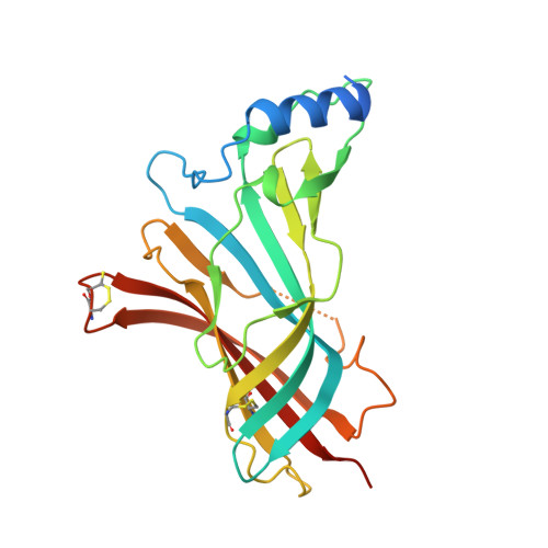
    Despite extensive studies on nicotinic acetylcholine receptors (nAChRs) and homologues, details of acetylcholine binding are not completely resolved. Here, we report the crystal structure of acetylcholine bound to the receptor homologue acetylcholine binding protein from Lymnaea stagnalis. This is the first structure of acetylcholine in a binding pocket containing all five aromatic residues conserved in all mammalian nAChRs. The ligand-protein interactions are characterized by contacts to the aromatic box formed primarily by residues on the principal side of the intersubunit binding interface (residues Tyr89, Trp143 and Tyr185). Besides these interactions on the principal side, we observe a cation-π interaction between acetylcholine and Trp53 on the complementary side and a water-mediated hydrogen bond from acetylcholine to backbone atoms of Leu102 and Met114, both of importance for anchoring acetylcholine to the complementary side. To further study the role of Trp53, we mutated the corresponding tryptophan in the two different acetylcholine-binding interfaces of the widespread α4β2 nAChR, i.e. the interfaces α4(+)β2(-) and α4(+)α4(-). Mutation to alanine (W82A on the β2 subunit or W88A on the α4 subunit) significantly altered the response to acetylcholine measured by oocyte voltage-clamp electrophysiology in both interfaces. This shows that the conserved tryptophan residue is important for the effects of ACh at α4β2 nAChRs, as also indicated by the crystal structure. The results add important details to the understanding of how this neurotransmitter exerts its action and improves the foundation for rational drug design targeting these receptors.


  • Organizational Affiliation

    Department of Drug Design and Pharmacology, Faculty of Health and Medical Sciences, University of Copenhagen, Copenhagen, Denmark; NeuroSearch A/S, Ballerup, Denmark.


Macromolecules
Find similar proteins by:  (by identity cutoff)  |  3D Structure
Entity ID: 1
MoleculeChains Sequence LengthOrganismDetailsImage
Acetylcholine-binding protein
A, B, C, D, E
A, B, C, D, E, F, G, H, I, J
229Lymnaea stagnalisMutation(s): 0 
UniProt
Find proteins for P58154 (Lymnaea stagnalis)
Explore P58154 
Go to UniProtKB:  P58154
Entity Groups  
Sequence Clusters30% Identity50% Identity70% Identity90% Identity95% Identity100% Identity
UniProt GroupP58154
Sequence Annotations
Expand
  • Reference Sequence
Small Molecules
Ligands 5 Unique
IDChains Name / Formula / InChI Key2D Diagram3D Interactions
1PE
Query on 1PE

Download Ideal Coordinates CCD File 
BA [auth E]
GA [auth G]
L [auth A]
LA [auth H]
M [auth A]
BA [auth E],
GA [auth G],
L [auth A],
LA [auth H],
M [auth A],
N [auth A],
NA [auth I],
Q [auth B],
QA [auth J],
R [auth B],
X [auth D],
Y [auth D]
PENTAETHYLENE GLYCOL
C10 H22 O6
JLFNLZLINWHATN-UHFFFAOYSA-N
ACH
Query on ACH

Download Ideal Coordinates CCD File 
CA [auth F]
EA [auth G]
K [auth A]
KA [auth H]
MA [auth I]
CA [auth F],
EA [auth G],
K [auth A],
KA [auth H],
MA [auth I],
O [auth B],
OA [auth J],
T [auth C],
V [auth D],
Z [auth E]
ACETYLCHOLINE
C7 H16 N O2
OIPILFWXSMYKGL-UHFFFAOYSA-N
SO4
Query on SO4

Download Ideal Coordinates CCD File 
AA [auth E]
DA [auth F]
FA [auth G]
P [auth B]
PA [auth J]
AA [auth E],
DA [auth F],
FA [auth G],
P [auth B],
PA [auth J],
U [auth C],
W [auth D]
SULFATE ION
O4 S
QAOWNCQODCNURD-UHFFFAOYSA-L
GOL
Query on GOL

Download Ideal Coordinates CCD File 
HA [auth G]GLYCEROL
C3 H8 O3
PEDCQBHIVMGVHV-UHFFFAOYSA-N
ACT
Query on ACT

Download Ideal Coordinates CCD File 
IA [auth G],
JA [auth G],
S [auth B]
ACETATE ION
C2 H3 O2
QTBSBXVTEAMEQO-UHFFFAOYSA-M
Binding Affinity Annotations 
IDSourceBinding Affinity
ACH BindingDB:  3WIP Ki: min: 3162.28, max: 6309.57 (nM) from 2 assay(s)
Kd: min: 458, max: 3850 (nM) from 2 assay(s)
Experimental Data & Validation

Experimental Data

  • Method: X-RAY DIFFRACTION
  • Resolution: 2.60 Å
  • R-Value Free: 0.239 
  • R-Value Work: 0.179 
  • R-Value Observed: 0.182 
  • Space Group: C 1 2 1
Unit Cell:
Length ( Å )Angle ( ˚ )
a = 236.85α = 90
b = 73.15β = 101.45
c = 132.47γ = 90
Software Package:
Software NamePurpose
SCALAdata scaling
PHENIXrefinement
PDB_EXTRACTdata extraction
MxCuBEdata collection
XSCALEdata scaling
PHASERphasing

Structure Validation

View Full Validation Report


Entry History 

Deposition Data

Revision History  (Full details and data files)

  • Version 1.0: 2014-04-23
    Type: Initial release
  • Version 1.1: 2018-05-16
    Changes: Data collection, Refinement description
  • Version 1.2: 2023-11-08
    Changes: Data collection, Database references, Derived calculations, Refinement description