3WFN

Crystal Structure of Nav1.6 IQ motif in complex with apo-CaM


Experimental Data Snapshot

  • Method: X-RAY DIFFRACTION
  • Resolution: 1.95 Å
  • R-Value Free: 0.237 
  • R-Value Work: 0.200 
  • R-Value Observed: 0.201 

wwPDB Validation   3D Report Full Report


This is version 1.2 of the entry. See complete history


Literature

Structural Basis for the Modulation of the Neuronal Voltage-Gated Sodium Channel NaV1.6 by Calmodulin

Chichili, V.P.R.Xiao, Y.Seetharaman, J.Cummins, T.R.Sivaraman, J.

(2013) Sci Rep 3: 2435-2435

  • DOI: https://doi.org/10.1038/srep02435
  • Primary Citation of Related Structures:  
    3WFN

  • PubMed Abstract: 

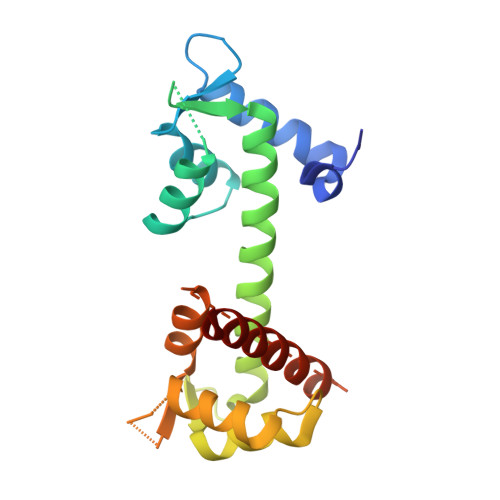
    The neuronal-voltage gated sodium channel (VGSC), Na(V)1.6, plays an important role in propagating action potentials along myelinated axons. Calmodulin (CaM) is known to modulate the inactivation kinetics of Na(V)1.6 by interacting with its IQ motif. Here we report the crystal structure of apo-CaM:Na(V)1.6IQ motif, along with functional studies. The IQ motif of Na(V)1.6 adopts an α-helical conformation in its interaction with the C-lobe of CaM. CaM uses different residues to interact with Na(V)1.6IQ motif depending on the presence or absence of Ca²⁺. Three residues from Na(V)1.6, Arg1902, Tyr1904 and Arg1905 were identified as the key common interacting residues in both the presence and absence of Ca²⁺. Substitution of Arg1902 and Tyr1904 with alanine showed a reduced rate of Na(V)1.6 inactivation in electrophysiological experiments in vivo. Compared with other CaM:Na(V) complexes, our results reveal a different mode of interaction for CaM:Na(V)1.6 and provides structural insight into the isoform-specific modulation of VGSCs.


  • Organizational Affiliation

    Department of Biological Sciences, National University of Singapore, Singapore, 117543.


Macromolecules
Find similar proteins by:  (by identity cutoff)  |  3D Structure
Entity ID: 1
MoleculeChains Sequence LengthOrganismDetailsImage
Calmodulin, Sodium channel protein type 8 subunit alphaA [auth B],
B [auth C],
C [auth D],
D [auth E]
182Mus musculusMutation(s): 0 
UniProt & NIH Common Fund Data Resources
Find proteins for Q9WTU3 (Mus musculus)
Explore Q9WTU3 
Go to UniProtKB:  Q9WTU3
IMPC:  MGI:103169
Find proteins for P0DP26 (Mus musculus)
Explore P0DP26 
Go to UniProtKB:  P0DP26
Entity Groups  
Sequence Clusters30% Identity50% Identity70% Identity90% Identity95% Identity100% Identity
UniProt GroupsQ9WTU3P0DP26
Sequence Annotations
Expand
  • Reference Sequence
Experimental Data & Validation

Experimental Data

  • Method: X-RAY DIFFRACTION
  • Resolution: 1.95 Å
  • R-Value Free: 0.237 
  • R-Value Work: 0.200 
  • R-Value Observed: 0.201 
  • Space Group: P 1 21 1
Unit Cell:
Length ( Å )Angle ( ˚ )
a = 72.83α = 90
b = 50.752β = 91.48
c = 150.666γ = 90
Software Package:
Software NamePurpose
ADSCdata collection
PHENIXmodel building
PHENIXrefinement
HKL-2000data reduction
HKL-2000data scaling
PHENIXphasing

Structure Validation

View Full Validation Report


Entry History 

Deposition Data

Revision History  (Full details and data files)

  • Version 1.0: 2013-08-28
    Type: Initial release
  • Version 1.1: 2017-08-16
    Changes: Source and taxonomy
  • Version 1.2: 2024-03-20
    Changes: Data collection, Database references