3WFJ

The complex structure of D-mandelate dehydrogenase with NADH


Experimental Data Snapshot

  • Method: X-RAY DIFFRACTION
  • Resolution: 2.80 Å
  • R-Value Free: 0.306 
  • R-Value Work: 0.239 
  • R-Value Observed: 0.242 

wwPDB Validation   3D Report Full Report


Ligand Structure Quality Assessment 


This is version 1.1 of the entry. See complete history


Literature

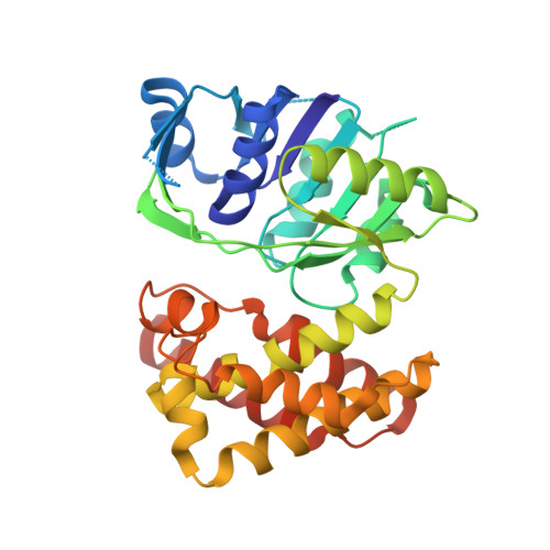
The crystal structure of D-mandelate dehydrogenase reveals its distinct substrate and coenzyme recognition mechanisms from those of 2-ketopantoate reductase.

Miyanaga, A.Fujisawa, S.Furukawa, N.Arai, K.Nakajima, M.Taguchi, H.

(2013) Biochem Biophys Res Commun 439: 109-114

  • DOI: https://doi.org/10.1016/j.bbrc.2013.08.019
  • Primary Citation of Related Structures:  
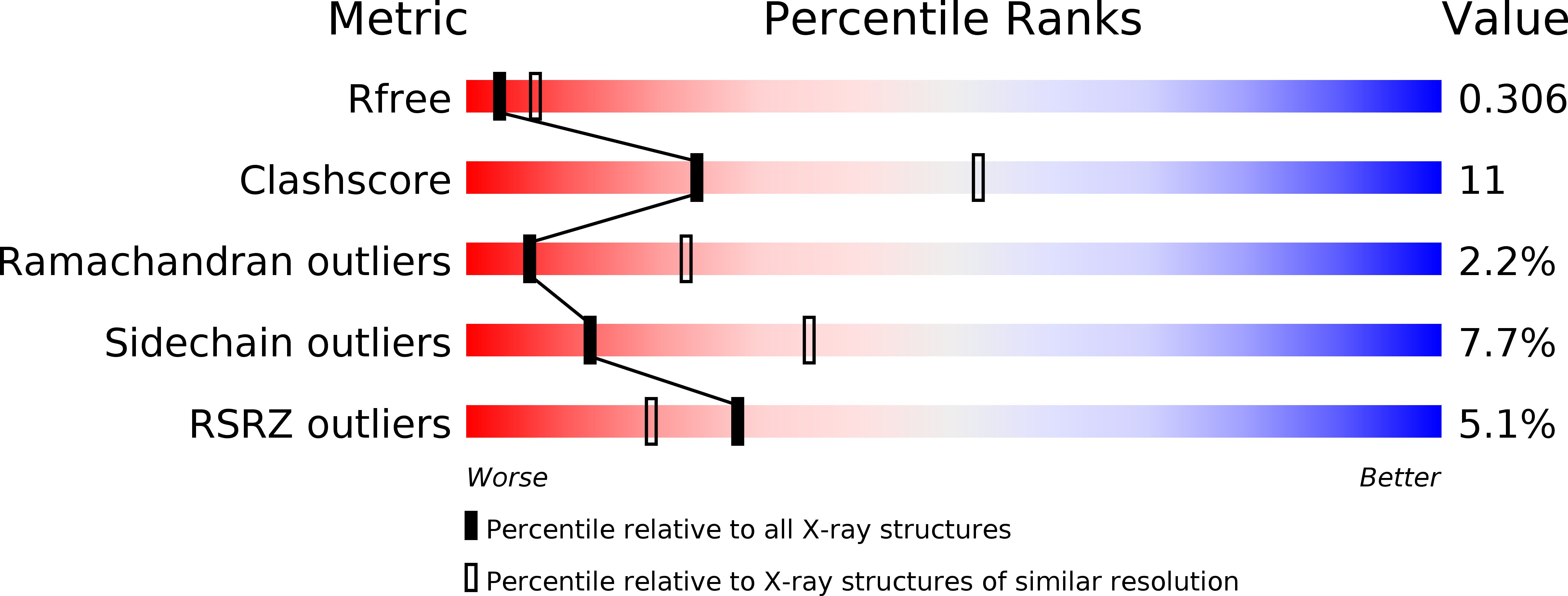
    3WFI, 3WFJ

  • PubMed Abstract: 

    D-Mandelate dehydrogenases (D-ManDHs), belonging to a new d-2-hydroxyacid dehydrogenase family, catalyze the conversion between benzoylformate and d-mandelate using NAD as a coenzyme. We determined the first D-ManDH structure, that of ManDH2 from Enterococcus faecalis IAM10071. The overall structure showed ManDH2 has a similar fold to 2-ketopantoate reductase (KPR), which catalyzes the conversion of 2-ketopantoate to d-pantoate using NADP as a coenzyme. They share conserved catalytic residues, indicating ManDH2 has the same reaction mechanism as KPR. However, ManDH2 exhibits significant structural variations in the coenzyme and substrate binding sites compared to KPR. These structural observations could explain their different coenzyme and substrate specificities.


  • Organizational Affiliation

    Department of Applied Biological Science, Faculty of Science and Technology, Tokyo University of Science, 2641 Yamazaki, Noda, Chiba 278-8510, Japan. Electronic address: miyanaga.a.aa@m.titech.ac.jp.


Macromolecules
Find similar proteins by:  (by identity cutoff)  |  3D Structure
Entity ID: 1
MoleculeChains Sequence LengthOrganismDetailsImage
2-dehydropantoate 2-reductase
A, B, C, D, E
A, B, C, D, E, F, G, H
312Enterococcus faeciumMutation(s): 0 
Gene Names: pLG1-0150
EC: 1.1.1.169
UniProt
Find proteins for E3USM3 (Enterococcus faecium)
Explore E3USM3 
Go to UniProtKB:  E3USM3
Entity Groups  
Sequence Clusters30% Identity50% Identity70% Identity90% Identity95% Identity100% Identity
UniProt GroupE3USM3
Sequence Annotations
Expand
  • Reference Sequence
Experimental Data & Validation

Experimental Data

  • Method: X-RAY DIFFRACTION
  • Resolution: 2.80 Å
  • R-Value Free: 0.306 
  • R-Value Work: 0.239 
  • R-Value Observed: 0.242 
  • Space Group: P 1 21 1
Unit Cell:
Length ( Å )Angle ( ˚ )
a = 115.65α = 90
b = 103.44β = 90.01
c = 119.74γ = 90
Software Package:
Software NamePurpose
ADSCdata collection
MOLREPphasing
REFMACrefinement
MOSFLMdata reduction
SCALAdata scaling

Structure Validation

View Full Validation Report


Ligand Structure Quality Assessment 


Entry History 

Deposition Data

Revision History  (Full details and data files)

  • Version 1.0: 2014-07-23
    Type: Initial release
  • Version 1.1: 2023-11-08
    Changes: Data collection, Database references, Derived calculations, Refinement description