3WCU

The structure of a deoxygenated 400 kda hemoglobin provides a more accurate description of the cooperative mechanism of giant hemoglobins: Deoxygenated form


Experimental Data Snapshot

  • Method: X-RAY DIFFRACTION
  • Resolution: 2.90 Å
  • R-Value Free: 0.292 
  • R-Value Work: 0.246 
  • R-Value Observed: 0.246 

wwPDB Validation   3D Report Full Report


Ligand Structure Quality Assessment 


This is version 2.1 of the entry. See complete history


Literature

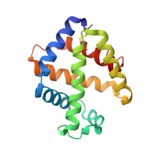
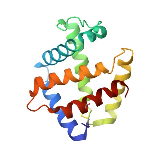
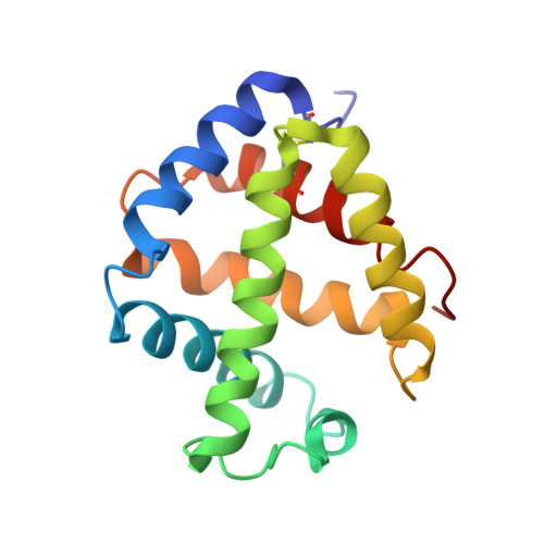
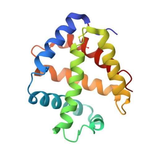
The structure of a deoxygenated 400 kDa haemoglobin reveals ternary- and quaternary-structural changes of giant haemoglobins

Numoto, N.Nakagawa, T.Ohara, R.Hasegawa, T.Kita, A.Yoshida, T.Maruyama, T.Imai, K.Fukumori, Y.Miki, K.

(2014) Acta Crystallogr D Biol Crystallogr 70: 1823-1831

  • DOI: https://doi.org/10.1107/S1399004714008475
  • Primary Citation of Related Structures:  
    3WCT, 3WCU, 3WCV, 3WCW

  • PubMed Abstract: 

    The quaternary structures of invertebrate haemoglobins (Hbs) are quite different from those of vertebrate Hbs. The extracellular giant Hbs of molecular masses of about 400 and 3600 kDa are composed of a dome-shaped dodecameric subassembly which consists of four individual globin subunits. Several crystal structures of 400 kDa Hbs from annelids have been reported, including structures in oxygenated and partially unliganded states, but the structure of the fully deoxygenated state has not been reported. In the present study, crystal structures of V2Hb from the tube worm Lamellibrachia satsuma have been determined in both the fully oxygenated and deoxygenated states. A glycosylation site and novel metal-binding sites for divalent cations were clearly observed with no intersubunit interactions in V2Hb. A comparison of the oxygenated and the deoxygenated forms of V2Hb reveals that the ternary- and quaternary-structural changes occur in a manner that maintains the molecular D3 symmetry. These structures suggest that the mechanisms of quaternary-structural changes between the oxy and deoxy states for the giant Hbs are identical across species.


  • Organizational Affiliation

    Department of Life Science, Graduate School of Natural Science and Technology, Kanazawa University, Kanazawa, Ishikawa 920-1192, Japan.


Macromolecules
Find similar proteins by:  (by identity cutoff)  |  3D Structure
Entity ID: 1
MoleculeChains Sequence LengthOrganismDetailsImage
A1 globin chain of giant V2 hemoglobin
A, E
146Lamellibrachia satsumaMutation(s): 0 
UniProt
Find proteins for S0BBU7 (Lamellibrachia satsuma)
Explore S0BBU7 
Go to UniProtKB:  S0BBU7
Entity Groups  
Sequence Clusters30% Identity50% Identity70% Identity90% Identity95% Identity100% Identity
UniProt GroupS0BBU7
Sequence Annotations
Expand
  • Reference Sequence
Find similar proteins by:  (by identity cutoff)  |  3D Structure
Entity ID: 2
MoleculeChains Sequence LengthOrganismDetailsImage
A2 globin chain of giant V2 hemoglobin
B, F
144Lamellibrachia satsumaMutation(s): 0 
UniProt
Find proteins for S0BBR6 (Lamellibrachia satsuma)
Explore S0BBR6 
Go to UniProtKB:  S0BBR6
Entity Groups  
Sequence Clusters30% Identity50% Identity70% Identity90% Identity95% Identity100% Identity
UniProt GroupS0BBR6
Sequence Annotations
Expand
  • Reference Sequence
Find similar proteins by:  (by identity cutoff)  |  3D Structure
Entity ID: 3
MoleculeChains Sequence LengthOrganismDetailsImage
B2 globin chain of giant V2 hemoglobin
C, G
150Lamellibrachia satsumaMutation(s): 0 
UniProt
Find proteins for S0BCU7 (Lamellibrachia satsuma)
Explore S0BCU7 
Go to UniProtKB:  S0BCU7
Entity Groups  
Sequence Clusters30% Identity50% Identity70% Identity90% Identity95% Identity100% Identity
UniProt GroupS0BCU7
Sequence Annotations
Expand
  • Reference Sequence
Find similar proteins by:  (by identity cutoff)  |  3D Structure
Entity ID: 4
MoleculeChains Sequence LengthOrganismDetailsImage
B1 globin chain of giant V2 hemoglobin
D, H
149Lamellibrachia satsumaMutation(s): 0 
UniProt
Find proteins for S0BAP9 (Lamellibrachia satsuma)
Explore S0BAP9 
Go to UniProtKB:  S0BAP9
Entity Groups  
Sequence Clusters30% Identity50% Identity70% Identity90% Identity95% Identity100% Identity
UniProt GroupS0BAP9
Sequence Annotations
Expand
  • Reference Sequence
Oligosaccharides

Help

Entity ID: 5
MoleculeChains Length2D Diagram Glycosylation3D Interactions
alpha-D-mannopyranose-(1-3)-[alpha-D-mannopyranose-(1-6)]alpha-D-mannopyranose-(1-4)-2-acetamido-2-deoxy-beta-D-glucopyranose-(1-4)-[alpha-L-fucopyranose-(1-6)]2-acetamido-2-deoxy-beta-D-glucopyranose
I, J
6N-Glycosylation
Glycosylation Resources
GlyTouCan:  G64181RO
GlyCosmos:  G64181RO
GlyGen:  G64181RO
Small Molecules
Ligands 2 Unique
IDChains Name / Formula / InChI Key2D Diagram3D Interactions
HEM
Query on HEM

Download Ideal Coordinates CCD File 
K [auth A]
L [auth B]
N [auth C]
O [auth D]
P [auth E]
K [auth A],
L [auth B],
N [auth C],
O [auth D],
P [auth E],
Q [auth F],
S [auth G],
T [auth H]
PROTOPORPHYRIN IX CONTAINING FE
C34 H32 Fe N4 O4
KABFMIBPWCXCRK-RGGAHWMASA-L
CA
Query on CA

Download Ideal Coordinates CCD File 
M [auth B],
R [auth F]
CALCIUM ION
Ca
BHPQYMZQTOCNFJ-UHFFFAOYSA-N
Experimental Data & Validation

Experimental Data

  • Method: X-RAY DIFFRACTION
  • Resolution: 2.90 Å
  • R-Value Free: 0.292 
  • R-Value Work: 0.246 
  • R-Value Observed: 0.246 
  • Space Group: P 63
Unit Cell:
Length ( Å )Angle ( ˚ )
a = 108.872α = 90
b = 108.872β = 90
c = 195.157γ = 120
Software Package:
Software NamePurpose
BSSdata collection
CNSrefinement
HKL-2000data reduction
HKL-2000data scaling
CNSphasing

Structure Validation

View Full Validation Report


Ligand Structure Quality Assessment 


Entry History 

Deposition Data

Revision History  (Full details and data files)

  • Version 1.0: 2014-06-04
    Type: Initial release
  • Version 1.1: 2014-08-06
    Changes: Database references
  • Version 2.0: 2020-07-29
    Type: Remediation
    Reason: Carbohydrate remediation
    Changes: Atomic model, Data collection, Derived calculations, Structure summary
  • Version 2.1: 2023-11-08
    Changes: Data collection, Database references, Refinement description, Structure summary