


3WAY
Crystal Structure of Autotaxin in Complex with 4BoA
- PDB DOI: https://doi.org/10.2210/pdb3WAY/pdb
- Classification: HYDROLASE/HYDROLASE INHIBITOR
- Organism(s): Mus musculus
- Expression System: Homo sapiens
- Mutation(s): No
- Deposited: 2013-05-09 Released: 2013-07-31
Experimental Data Snapshot
- Method: X-RAY DIFFRACTION
- Resolution: 1.75 Å
- R-Value Free: 0.221
- R-Value Work: 0.181
- R-Value Observed: 0.183

This is version 2.1 of the entry. See complete history.

Macromolecules
Find similar proteins by:
(by identity cutoff) | 3D Structure
Entity ID: 1 | |||||
|---|---|---|---|---|---|
| Molecule | Chains | Sequence Length | Organism | Details | Image |
| Ectonucleotide pyrophosphatase/phosphodiesterase family member 2 | 831 | Mus musculus | Mutation(s): 0 Gene Names: Enpp2 EC: 3.1.4.39 | 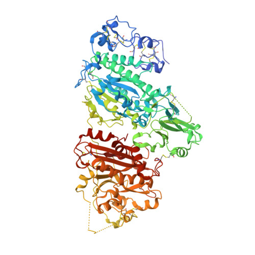 | |
UniProt | |||||
Find proteins for Q9R1E6 (Mus musculus) Explore Q9R1E6 Go to UniProtKB: Q9R1E6 | |||||
Entity Groups | |||||
| Sequence Clusters | 30% Identity50% Identity70% Identity90% Identity95% Identity100% Identity | ||||
| UniProt Group | Q9R1E6 | ||||
Sequence AnnotationsExpand | |||||
| |||||
Oligosaccharides
Entity ID: 2 | |||||
|---|---|---|---|---|---|
| Molecule | Chains | Length | 2D Diagram | Glycosylation | 3D Interactions |
| 2-acetamido-2-deoxy-beta-D-glucopyranose-(1-4)-2-acetamido-2-deoxy-beta-D-glucopyranose | B, D | 2 |  | N-Glycosylation | |
Glycosylation Resources | |||||
GlyTouCan: G42666HT GlyCosmos: G42666HT GlyGen: G42666HT | |||||
Entity ID: 3 | |||||
|---|---|---|---|---|---|
| Molecule | Chains | Length | 2D Diagram | Glycosylation | 3D Interactions |
| alpha-D-mannopyranose-(1-2)-alpha-D-mannopyranose-(1-3)-alpha-D-mannopyranose-(1-6)-beta-D-mannopyranose-(1-4)-2-acetamido-2-deoxy-beta-D-glucopyranose-(1-4)-2-acetamido-2-deoxy-beta-D-glucopyranose | C | 6 |  | N-Glycosylation | |
Glycosylation Resources | |||||
GlyTouCan: G47410OF GlyCosmos: G47410OF GlyGen: G47410OF | |||||
Small Molecules
| Ligands 7 Unique | |||||
|---|---|---|---|---|---|
| ID | Chains | Name / Formula / InChI Key | 2D Diagram | 3D Interactions | |
| DWY Query on DWY | Y [auth A] | [4-({4-[(5Z)-5-(3,4-dichlorobenzylidene)-4-oxo-4,5-dihydro-1,3-thiazol-2-yl]piperazin-1-yl}methyl)phenyl]boronic acid C21 H20 B Cl2 N3 O3 S CKUAZJLGUCZSNN-UNOMPAQXSA-N |  | ||
| ZN Query on ZN | E [auth A], F [auth A] | ZINC ION Zn PTFCDOFLOPIGGS-UHFFFAOYSA-N |  | ||
| EDO Query on EDO | M [auth A] N [auth A] O [auth A] P [auth A] Q [auth A] | 1,2-ETHANEDIOL C2 H6 O2 LYCAIKOWRPUZTN-UHFFFAOYSA-N |  | ||
| SCN Query on SCN | J [auth A], K [auth A], L [auth A] | THIOCYANATE ION C N S ZMZDMBWJUHKJPS-UHFFFAOYSA-M |  | ||
| CA Query on CA | G [auth A] | CALCIUM ION Ca BHPQYMZQTOCNFJ-UHFFFAOYSA-N |  | ||
| K Query on K | I [auth A] | POTASSIUM ION K NPYPAHLBTDXSSS-UHFFFAOYSA-N |  | ||
| NA Query on NA | H [auth A] | SODIUM ION Na FKNQFGJONOIPTF-UHFFFAOYSA-N |  | ||
Experimental Data & Validation
Experimental Data
- Method: X-RAY DIFFRACTION
- Resolution: 1.75 Å
- R-Value Free: 0.221
- R-Value Work: 0.181
- R-Value Observed: 0.183
- Space Group: P 1 21 1
Unit Cell:
| Length ( Å ) | Angle ( ˚ ) |
|---|---|
| a = 61.556 | α = 90 |
| b = 94.524 | β = 93.98 |
| c = 75.242 | γ = 90 |
| Software Name | Purpose |
|---|---|
| PHENIX | refinement |
| PDB_EXTRACT | data extraction |
| HKL-2000 | data collection |
| HKL-2000 | data reduction |
| HKL-2000 | data scaling |
| MOLREP | phasing |
Entry History
Deposition Data
- Released Date: 2013-07-31 Deposition Author(s): Nishimasu, H., Ishitani, R., Nureki, O.
Revision History (Full details and data files)
- Version 1.0: 2013-07-31
Type: Initial release - Version 1.1: 2013-10-16
Changes: Database references - Version 2.0: 2020-07-29
Type: Remediation
Reason: Carbohydrate remediation
Changes: Atomic model, Data collection, Database references, Derived calculations, Structure summary - Version 2.1: 2023-11-08
Changes: Data collection, Database references, Refinement description, Structure summary
