3W45

Crystal structure of RsbX in complex with cobalt in space group P1


Experimental Data Snapshot

  • Method: X-RAY DIFFRACTION
  • Resolution: 1.70 Å
  • R-Value Free: 0.202 
  • R-Value Work: 0.170 
  • R-Value Observed: 0.172 

wwPDB Validation   3D Report Full Report


This is version 1.2 of the entry. See complete history


Literature

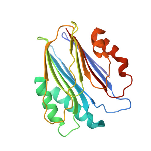
Structure of the RsbX phosphatase involved in the general stress response of Bacillus subtilis

Teh, A.H.Makino, M.Hoshino, T.Baba, S.Shimizu, N.Yamamoto, M.Kumasaka, T.

(2015) Acta Crystallogr D Biol Crystallogr 71: 1392-1399

  • DOI: https://doi.org/10.1107/S1399004715007166
  • Primary Citation of Related Structures:  
    3W40, 3W41, 3W42, 3W43, 3W44, 3W45

  • PubMed Abstract: 

    In the general stress response of Bacillus subtilis, which is governed by the sigma factor σ(B), stress signalling is relayed by a cascade of Rsb proteins that regulate σ(B) activity. RsbX, a PPM II phosphatase, halts the response by dephosphorylating the stressosome composed of RsbR and RsbS. The crystal structure of RsbX reveals a reorganization of the catalytic centre, with the second Mn(2+) ion uniquely coordinated by Gly47 O from the β4-α1 loop instead of a water molecule as in PPM I phosphatases. An extra helical turn of α1 tilts the loop towards the metal-binding site, and the β2-β3 loop swings outwards to accommodate this tilting. The residues critical for this defining feature of the PPM II phosphatases are highly conserved. Formation of the catalytic centre is metal-specific, as crystallization with Mg(2+) ions resulted in a shift of the β4-α1 loop that led to loss of the second ion. RsbX also lacks the flap subdomain characteristic of PPM I phosphatases. On the basis of a stressosome model, the activity of RsbX towards RsbR-P and RsbS-P may be influenced by the different accessibilities of their phosphorylation sites.


  • Organizational Affiliation

    Japan Synchrotron Radiation Research Institute (JASRI/SPring-8), 1-1-1 Kouto, Sayo, Hyogo 679-5198, Japan.


Macromolecules
Find similar proteins by:  (by identity cutoff)  |  3D Structure
Entity ID: 1
MoleculeChains Sequence LengthOrganismDetailsImage
Phosphoserine phosphatase RsbX
A, B
199Bacillus subtilis subsp. subtilis str. 168Mutation(s): 0 
Gene Names: BSU04740rsbX
EC: 3.1.3.3
UniProt
Find proteins for P17906 (Bacillus subtilis (strain 168))
Explore P17906 
Go to UniProtKB:  P17906
Entity Groups  
Sequence Clusters30% Identity50% Identity70% Identity90% Identity95% Identity100% Identity
UniProt GroupP17906
Sequence Annotations
Expand
  • Reference Sequence
Experimental Data & Validation

Experimental Data

  • Method: X-RAY DIFFRACTION
  • Resolution: 1.70 Å
  • R-Value Free: 0.202 
  • R-Value Work: 0.170 
  • R-Value Observed: 0.172 
  • Space Group: P 1
Unit Cell:
Length ( Å )Angle ( ˚ )
a = 33.108α = 80.81
b = 41.614β = 89.95
c = 68.806γ = 71.08
Software Package:
Software NamePurpose
REFMACrefinement
PDB_EXTRACTdata extraction
BSSdata collection
HKL-2000data reduction
HKL-2000data scaling
SHELXSphasing

Structure Validation

View Full Validation Report


Entry History 

Deposition Data

Revision History  (Full details and data files)

  • Version 1.0: 2014-01-22
    Type: Initial release
  • Version 1.1: 2015-07-01
    Changes: Database references
  • Version 1.2: 2024-03-20
    Changes: Data collection, Database references, Derived calculations