Identification and structural characterization of a new three-finger toxin hemachatoxin from Hemachatus haemachatus venom.
Girish, V.M., Kumar, S., Joseph, L., Jobichen, C., Kini, R.M., Sivaraman, J.(2012) PLoS One 7: e48112-e48112
- PubMed: 23144733
- DOI: https://doi.org/10.1371/journal.pone.0048112
- Primary Citation of Related Structures:
3VTS - PubMed Abstract:
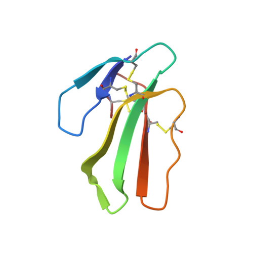
Snake venoms are rich sources of biologically active proteins and polypeptides. Three-finger toxins are non-enzymatic proteins present in elapid (cobras, kraits, mambas and sea snakes) and colubrid venoms. These proteins contain four conserved disulfide bonds in the core to maintain the three-finger folds. Although all three-finger toxins have similar fold, their biological activities are different. A new three-finger toxin (hemachatoxin) was isolated from Hemachatus haemachatus (Ringhals cobra) venom. Its amino acid sequence was elucidated, and crystal structure was determined at 2.43 Å resolution. The overall fold is similar to other three-finger toxins. The structure and sequence analysis revealed that the fold is maintained by four highly conserved disulfide bonds. It exhibited highest similarity to particularly P-type cardiotoxins that are known to associate and perturb the membrane surface with their lipid binding sites. Also, the increased B value of hemachotoxin loop II suggests that loop II is flexible and may remain flexible until its interaction with membrane phospholipids. Based on the analysis, we predict hemachatoxin to be cardiotoxic/cytotoxic and our future experiments will be directed to characterize the activity of hemachatoxin.
Organizational Affiliation:
Department of Biological Sciences, Faculty of Science, National University of Singapore, Singapore, Singapore.