3VSR

Microbacterium saccharophilum K-1 beta-fructofuranosidase catalytic domain


Experimental Data Snapshot

  • Method: X-RAY DIFFRACTION
  • Resolution: 2.00 Å
  • R-Value Free: 0.241 
  • R-Value Work: 0.193 
  • R-Value Observed: 0.196 

wwPDB Validation   3D Report Full Report


This is version 1.4 of the entry. See complete history


Literature

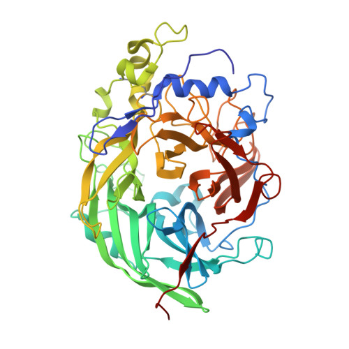
Crystal structure of a lactosucrose-producing enzyme, Arthrobacter sp. K-1 beta-fructofuranosidase

Tonozuka, T.Tamaki, A.Yokoi, G.Miyazaki, T.Ichikawa, M.Nishikawa, A.Ohta, Y.Hidaka, Y.Katayama, K.Hatada, Y.Ito, T.Fujita, K.

(2012) Enzyme Microb Technol 51: 359-365

  • DOI: https://doi.org/10.1016/j.enzmictec.2012.08.004
  • Primary Citation of Related Structures:  
    3VSR

  • PubMed Abstract: 

    Arthrobacter sp. K-1 β-fructofuranosidase (ArFFase), a glycoside hydrolase family 68 enzyme, catalyzes the hydrolysis and transfructosylation of sucrose. ArFFase is useful for producing a sweetener, lactosucrose (4(G)-β-D-galactosylsucrose). The primary structure of ArFFase is homologous to those of levansucrases, although ArFFase catalyzes mostly hydrolysis when incubated with sucrose alone, even at high concentration. Here, we determined the crystal structure of ArFFase in unliganded form and complexed with fructose. ArFFase consisted of a five-bladed β-propeller fold as observed in levansucrases. The structure of ArFFase was most similar to that of Gluconacetobacter diazotrophicus levansucrase (GdLev). The structure of the catalytic cleft of ArFFase was also highly homologous to that of GdLev. However, two amino acid residues, Tyr232 and Pro442 in ArFFase, were not conserved between them. A tunnel observed at the bottom of the catalytic cleft of ArFFase may serve as a water drain or its reservoir.


  • Organizational Affiliation

    Department of Applied Biological Science, Tokyo University of Agriculture and Technology, 3-5-8 Saiwai-cho, Fuchu, Tokyo 186-0001, Japan. tonozuka@cc.tuat.ac.jp


Macromolecules
Find similar proteins by:  (by identity cutoff)  |  3D Structure
Entity ID: 1
MoleculeChains Sequence LengthOrganismDetailsImage
Beta-fructofuranosidase496Microbacterium saccharophilumMutation(s): 0 
Gene Names: bff
EC: 3.2.1.26
UniProt
Find proteins for Q8VW87 (Microbacterium saccharophilum)
Explore Q8VW87 
Go to UniProtKB:  Q8VW87
Entity Groups  
Sequence Clusters30% Identity50% Identity70% Identity90% Identity95% Identity100% Identity
UniProt GroupQ8VW87
Sequence Annotations
Expand
  • Reference Sequence
Experimental Data & Validation

Experimental Data

  • Method: X-RAY DIFFRACTION
  • Resolution: 2.00 Å
  • R-Value Free: 0.241 
  • R-Value Work: 0.193 
  • R-Value Observed: 0.196 
  • Space Group: P 1 21 1
Unit Cell:
Length ( Å )Angle ( ˚ )
a = 57.723α = 90
b = 72.391β = 95.23
c = 58.709γ = 90
Software Package:
Software NamePurpose
ADSCdata collection
MOLREPphasing
REFMACrefinement
HKL-2000data reduction
HKL-2000data scaling

Structure Validation

View Full Validation Report


Entry History 

Deposition Data

Revision History  (Full details and data files)

  • Version 1.0: 2012-08-22
    Type: Initial release
  • Version 1.1: 2012-10-17
    Changes: Database references
  • Version 1.2: 2012-10-31
    Changes: Database references
  • Version 1.3: 2014-03-12
    Changes: Structure summary
  • Version 1.4: 2023-11-08
    Changes: Data collection, Database references, Refinement description