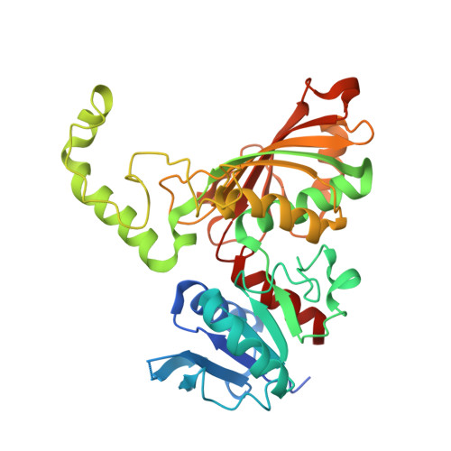
Structures of ternary complexes of aspartate-semialdehyde dehydrogenase (Rv3708c) from Mycobacterium tuberculosis H37Rv
Vyas, R., Tewari, R., Weiss, M.S., Karthikeyan, S.(2012) Acta Crystallogr D Biol Crystallogr 68: 671-679
- PubMed: 22683789
- DOI: https://doi.org/10.1107/S0907444912007330
- Primary Citation of Related Structures:
3TZ6, 3VOS - PubMed Abstract:
Aspartate-semialdehyde dehydrogenase (Asd; ASADH; EC 1.2.1.11) is the enzyme that lies at the first branch point in the biosynthetic pathway of important amino acids including lysine and methionine and the cell-wall component diaminopimelate (DAP). The enzymatic reaction of ASADH is the reductive dephosphorylation of aspartyl-β-phosphate (ABP) to aspartate β-semialdehyde (ASA). Since the aspartate pathway is absolutely essential for the survival of many microbes and is absent in humans, the enzymes involved in this pathway can be considered to be potential antibacterial drug targets. In this work, the structure of ASADH from Mycobacterium tuberculosis H37Rv (Mtb-ASADH) has been determined in complex with glycerol and sulfate at 2.18 Å resolution and in complex with S-methyl-L-cysteine sulfoxide (SMCS) and sulfate at 1.95 Å resolution. The overall structure of Mtb-ASADH is similar to those of its orthologues. However, in the Mtb-ASADH-glycerol complex structure the glycerol molecule is noncovalently bound to the active-site residue Cys130, while in the Mtb-ASADH-SMCS complex structure the SMCS (Cys) is covalently linked to Cys130. The Mtb-ASADH-SMCS complex structurally mimics one of the intermediate steps in the proposed mechanism of ASADH enzyme catalysis. Comparison of the two complex structures revealed that the amino acids Glu224 and Arg249 undergo conformational changes upon binding of glycerol. Moreover, the structures reported here may help in the development of species-specific antibacterial drug molecules against human pathogens.
Organizational Affiliation:
Department of Biotechnology, Panjab University, Chandigarh 160 014, India.