


Crystal structure of the human APOBEC3C having HIV-1 Vif-binding interface
Kitamura, S., Ode, H., Nakashima, M., Imahashi, M., Naganawa, Y., Ibe, S., Yokomaku, Y., Watanabe, N., Suzuki, A., Sugiura, W., Iwatani, Y.To be published.
Experimental Data Snapshot
wwPDB Validation 3D Report Full Report

Entity ID: 1 | |||||
|---|---|---|---|---|---|
| Molecule | Chains | Sequence Length | Organism | Details | Image |
| Probable DNA dC->dU-editing enzyme APOBEC-3C | 190 | Homo sapiens | Mutation(s): 0 Gene Names: APOBEC3C, APOBEC1L, PBI EC: 3.5.4 | 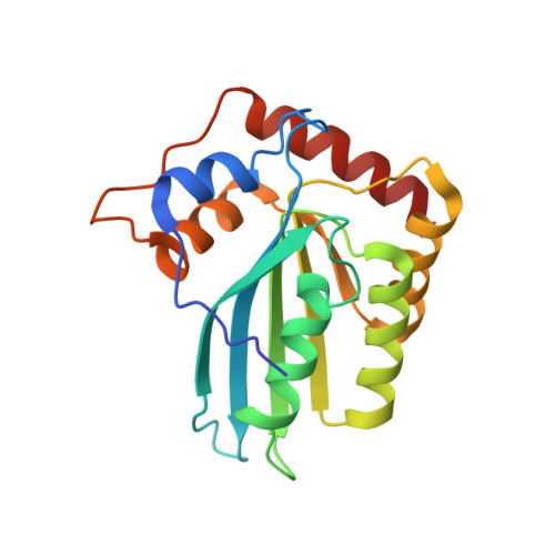 | |
UniProt & NIH Common Fund Data Resources | |||||
Find proteins for Q9NRW3 (Homo sapiens) Explore Q9NRW3 Go to UniProtKB: Q9NRW3 | |||||
PHAROS: Q9NRW3 GTEx: ENSG00000244509 | |||||
Entity Groups | |||||
| Sequence Clusters | 30% Identity50% Identity70% Identity90% Identity95% Identity100% Identity | ||||
| UniProt Group | Q9NRW3 | ||||
Sequence AnnotationsExpand | |||||
| |||||
| Ligands 1 Unique | |||||
|---|---|---|---|---|---|
| ID | Chains | Name / Formula / InChI Key | 2D Diagram | 3D Interactions | |
| ZN Query on ZN | C [auth A], D [auth B] | ZINC ION Zn PTFCDOFLOPIGGS-UHFFFAOYSA-N |  | ||
| Length ( Å ) | Angle ( ˚ ) |
|---|---|
| a = 104.419 | α = 90 |
| b = 104.419 | β = 90 |
| c = 74.502 | γ = 120 |
| Software Name | Purpose |
|---|---|
| SCALEPACK | data scaling |
| REFMAC | refinement |
| PDB_EXTRACT | data extraction |
| CrystalClear | data collection |
| HKL-2000 | data reduction |
| HKL-2000 | data scaling |
| MOLREP | phasing |
RCSB PDB (citation) is hosted by
RCSB PDB is a member of the

