3VG5

Barium derivative of human LFABP


Experimental Data Snapshot

  • Method: X-RAY DIFFRACTION
  • Resolution: 2.00 Å
  • R-Value Free: 0.250 
  • R-Value Work: 0.214 

wwPDB Validation   3D Report Full Report


Ligand Structure Quality Assessment 


This is version 1.2 of the entry. See complete history


Literature

Utility of anion and cation combinations for phasing of protein structures.

Sharma, A.Yogavel, M.Sharma, A.

(2012) J Struct Funct Genomics 13: 135-143

  • DOI: https://doi.org/10.1007/s10969-012-9137-3
  • Primary Citation of Related Structures:  
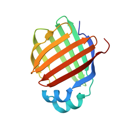
    3B2H, 3B2I, 3B2J, 3B2K, 3B2L, 3VG2, 3VG3, 3VG4, 3VG5, 3VG6, 3VG7

  • PubMed Abstract: 

    We report the use of anionic (I(-)), cationic (Ba(2+), Cd(2+)) and ionic mixtures (I(-) plus Ba(2+)) for derivatizing liver fatty acid binding protein (LFABP) crystals. Use of cationic and anionic salts in phasing experiments revealed distinct non-overlapping sites for these ions, suggesting exclusive binding regions on LFABP. Interestingly, cations of identical charge and valency (like Ba(2+) and Cd(2+)) bound to distinct pockets on the protein surface. Furthermore, a mixture of salts containing both I(-) and Ba(2+) was very useful in phasing experiments as these oppositely charged ions bound to different regions of LFABP. Our data therefore suggest that cationic and anionic salt mixtures like BaCl(2) with NH(4)I or salts like CdI, BaI where each ion has a significant anomalous signal for a given X-ray wavelength may be valuable reagents for phasing during structure determination.


  • Organizational Affiliation

    Structural and Computational Biology Group, International Centre for Genetic Engineering and Biotechnology, New Delhi, India.


Macromolecules
Find similar proteins by:  (by identity cutoff)  |  3D Structure
Entity ID: 1
MoleculeChains Sequence LengthOrganismDetailsImage
Fatty acid-binding protein, liver132Homo sapiensMutation(s): 0 
Gene Names: FABP1FABPL
UniProt & NIH Common Fund Data Resources
Find proteins for P07148 (Homo sapiens)
Explore P07148 
Go to UniProtKB:  P07148
PHAROS:  P07148
GTEx:  ENSG00000163586 
Entity Groups  
Sequence Clusters30% Identity50% Identity70% Identity90% Identity95% Identity100% Identity
UniProt GroupP07148
Sequence Annotations
Expand
  • Reference Sequence
Experimental Data & Validation

Experimental Data

  • Method: X-RAY DIFFRACTION
  • Resolution: 2.00 Å
  • R-Value Free: 0.250 
  • R-Value Work: 0.214 
  • Space Group: P 21 21 21
Unit Cell:
Length ( Å )Angle ( ˚ )
a = 30.293α = 90
b = 57.122β = 90
c = 74.11γ = 90
Software Package:
Software NamePurpose
SCALEPACKdata scaling
REFMACrefinement
PDB_EXTRACTdata extraction
MAR345dtbdata collection
HKL-2000data reduction
HKL-2000data scaling
PHENIXphasing

Structure Validation

View Full Validation Report


Ligand Structure Quality Assessment 


Entry History 

Deposition Data

Revision History  (Full details and data files)

  • Version 1.0: 2012-06-20
    Type: Initial release
  • Version 1.1: 2013-06-19
    Changes: Database references
  • Version 1.2: 2024-03-20
    Changes: Data collection, Database references, Derived calculations