


3V1W
Molecular Basis for Multiple Ligand Binding of Calsequestrin and Potential Inhibition by Caffeine and Gallocatecin
- PDB DOI: https://doi.org/10.2210/pdb3V1W/pdb
- Classification: CALCIUM BINDING PROTEIN
- Organism(s): Oryctolagus cuniculus
- Mutation(s): No
- Deposited: 2011-12-10 Released: 2012-12-12
Experimental Data Snapshot
- Method: X-RAY DIFFRACTION
- Resolution: 1.91 Å
- R-Value Free: 0.222
- R-Value Work: 0.171
- R-Value Observed: 0.174
wwPDB Validation 3D Report Full Report

This is version 2.1 of the entry. See complete history.

Macromolecules
Find similar proteins by:
(by identity cutoff) | 3D Structure
Entity ID: 1 | |||||
|---|---|---|---|---|---|
| Molecule | Chains | Sequence Length | Organism | Details | Image |
| Calsequestrin-1 | 367 | Oryctolagus cuniculus | Mutation(s): 0 | 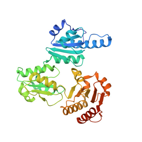 | |
UniProt | |||||
Find proteins for P07221 (Oryctolagus cuniculus) Explore P07221 Go to UniProtKB: P07221 | |||||
Entity Groups | |||||
| Sequence Clusters | 30% Identity50% Identity70% Identity90% Identity95% Identity100% Identity | ||||
| UniProt Group | P07221 | ||||
Sequence AnnotationsExpand | |||||
| |||||
Oligosaccharides

Small Molecules
| Ligands 4 Unique | |||||
|---|---|---|---|---|---|
| ID | Chains | Name / Formula / InChI Key | 2D Diagram | 3D Interactions | |
| MRD Query on MRD | E [auth A] | (4R)-2-METHYLPENTANE-2,4-DIOL C6 H14 O2 SVTBMSDMJJWYQN-RXMQYKEDSA-N |  | ||
| MPD Query on MPD | C [auth A], D [auth A] | (4S)-2-METHYL-2,4-PENTANEDIOL C6 H14 O2 SVTBMSDMJJWYQN-YFKPBYRVSA-N |  | ||
| CA Query on CA | F [auth A], G [auth A], H [auth A], I [auth A] | CALCIUM ION Ca BHPQYMZQTOCNFJ-UHFFFAOYSA-N |  | ||
| NA Query on NA | J [auth A], K [auth A] | SODIUM ION Na FKNQFGJONOIPTF-UHFFFAOYSA-N |  | ||
Experimental Data & Validation
Experimental Data
- Method: X-RAY DIFFRACTION
- Resolution: 1.91 Å
- R-Value Free: 0.222
- R-Value Work: 0.171
- R-Value Observed: 0.174
- Space Group: C 2 2 21
Unit Cell:
| Length ( Å ) | Angle ( ˚ ) |
|---|---|
| a = 59.082 | α = 90 |
| b = 144.592 | β = 90 |
| c = 110.965 | γ = 90 |
| Software Name | Purpose |
|---|---|
| PHENIX | refinement |
Entry History
Deposition Data
- Released Date: 2012-12-12 Deposition Author(s): Subramanian, A.K., Nissen, M.N., Lewis, K.M., Sanchez, E.J., Muralidharan, A.K., Kang, C.
Revision History (Full details and data files)
- Version 1.0: 2012-12-12
Type: Initial release - Version 2.0: 2020-07-29
Type: Remediation
Reason: Carbohydrate remediation
Changes: Advisory, Atomic model, Data collection, Derived calculations, Structure summary - Version 2.1: 2023-09-13
Changes: Advisory, Data collection, Database references, Refinement description, Structure summary
