X-ray crystallography and NMR studies of domain-swapped canecystatin-1.
Valadares, N.F., de Oliveira-Silva, R., Cavini, I.A., Marques, I.A., Pereira, H.D., Soares-Costa, A., Henrique-Silva, F., Kalbitzer, H.R., Munte, C.E., Garratt, R.C.(2013) FEBS J 280: 1028-1038
- PubMed: 23241243
- DOI: https://doi.org/10.1111/febs.12095
- Primary Citation of Related Structures:
3UL5, 3UL6 - PubMed Abstract:
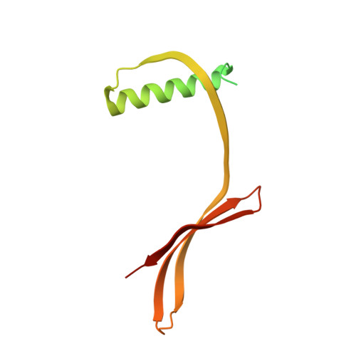
The three-dimensional structure of canecystatin-1, a potent inhibitor of cysteine proteases from sugarcane (Saccharum officinarum), has been solved in two different crystal forms. In both cases, it is seen to exist as a domain-swapped dimer, the first such observation for a cystatin of plant origin. Size exclusion chromatography and multidimensional NMR spectroscopy show the dimer to be the dominant species in solution, despite the presence of a measurable quantity of monomer undergoing slow exchange. The latter is believed to be the active species, whereas the domain-swapped dimer is presumably inactive, as its first inhibitory loop has been extended to form part of a long β-strand that forms a double-helical coiled coil with its partner from the other monomer. A similar structure is observed in human cystatin C, but the spatial disposition of the two lobes of the dimer is rather different. Dimerization is presumably a mechanism by which canecystatin-1 can be kept inactive within the plant, avoiding the inhibition of endogenous proteases. The structure described here provides a platform for the rational design of specific cysteine protease inhibitors for biotechnological applications.
Organizational Affiliation:
Center for Structural Molecular Biotechnology, Department of Physics and Informatics, Physics Institute of São Carlos, University of São Paulo, São Carlos-SP, Brazil.