3UKL

Crystal structure of UDP-galactopyranose mutase from Aspergillus fumigatus in complex with UDP


Experimental Data Snapshot

  • Method: X-RAY DIFFRACTION
  • Resolution: 2.63 Å
  • R-Value Free: 0.231 
  • R-Value Work: 0.185 
  • R-Value Observed: 0.187 

wwPDB Validation   3D Report Full Report


Ligand Structure Quality Assessment 


This is version 1.5 of the entry. See complete history


Literature

Structural Insight into the Unique Substrate Binding Mechanism and Flavin Redox State of UDP-galactopyranose Mutase from Aspergillus fumigatus.

van Straaten, K.E.Routier, F.H.Sanders, D.A.

(2012) J Biol Chem 287: 10780-10790

  • DOI: https://doi.org/10.1074/jbc.M111.322974
  • Primary Citation of Related Structures:  
    3UKA, 3UKF, 3UKH, 3UKK, 3UKL, 3UKP, 3UKQ

  • PubMed Abstract: 

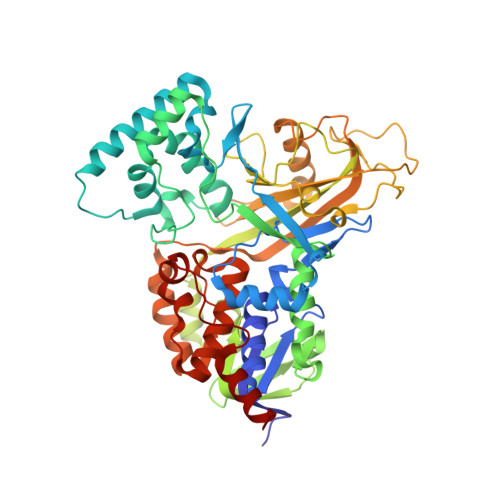
    UDP-galactopyranose mutase (UGM) is a flavin-containing enzyme that catalyzes the reversible conversion of UDP-galactopyranose (UDP-Galp) to UDP-galactofuranose (UDP-Galf). As in prokaryotic UGMs, the flavin needs to be reduced for the enzyme to be active. Here we present the first eukaryotic UGM structures from Aspergillus fumigatus (AfUGM). The structures are of UGM alone, with the substrate UDP-Galp and with the inhibitor UDP. Additionally, we report the structures of AfUGM bound to substrate with oxidized and reduced flavin. These structures provide insight into substrate recognition and structural changes observed upon substrate binding involving the mobile loops and the critical arginine residues Arg-182 and Arg-327. Comparison with prokaryotic UGM reveals that despite low sequence identity with known prokaryotic UGMs the overall fold is largely conserved. Structural differences between prokaryotic UGM and AfUGM result from inserts in AfUGM. A notable difference from prokaryotic UGMs is that AfUGM contains a third flexible loop (loop III) above the si-face of the isoalloxazine ring that changes position depending on the redox state of the flavin cofactor. This loop flipping has not been observed in prokaryotic UGMs. In addition we have determined the crystals structures and steady-state kinetic constants of the reaction catalyzed by mutants R182K, R327K, R182A, and R327A. These results support our hypothesis that Arg-182 and Arg-327 play important roles in stabilizing the position of the diphosphates of the nucleotide sugar and help to facilitate the positioning of the galactose moiety for catalysis.


  • Organizational Affiliation

    Department of Chemistry, University of Saskatchewan, Saskatoon, Saskatchewan S7N 5C9, Canada.


Macromolecules
Find similar proteins by:  (by identity cutoff)  |  3D Structure
Entity ID: 1
MoleculeChains Sequence LengthOrganismDetailsImage
UDP-galactopyranose mutase
A, B, C, D, E
A, B, C, D, E, F, G, H
510Aspergillus fumigatusMutation(s): 0 
Gene Names: glfglfA
EC: 5.4.99.9
UniProt
Find proteins for Q4W1X2 (Aspergillus fumigatus)
Explore Q4W1X2 
Go to UniProtKB:  Q4W1X2
Entity Groups  
Sequence Clusters30% Identity50% Identity70% Identity90% Identity95% Identity100% Identity
UniProt GroupQ4W1X2
Sequence Annotations
Expand
  • Reference Sequence
Small Molecules
Ligands 3 Unique
IDChains Name / Formula / InChI Key2D Diagram3D Interactions
FAD
Query on FAD

Download Ideal Coordinates CCD File 
CA [auth G]
EA [auth H]
J [auth A]
N [auth B]
Q [auth C]
CA [auth G],
EA [auth H],
J [auth A],
N [auth B],
Q [auth C],
T [auth D],
V [auth E],
Y [auth F]
FLAVIN-ADENINE DINUCLEOTIDE
C27 H33 N9 O15 P2
VWWQXMAJTJZDQX-UYBVJOGSSA-N
UDP
Query on UDP

Download Ideal Coordinates CCD File 
BA [auth G]
DA [auth H]
I [auth A]
M [auth B]
P [auth C]
BA [auth G],
DA [auth H],
I [auth A],
M [auth B],
P [auth C],
S [auth D],
U [auth E],
X [auth F]
URIDINE-5'-DIPHOSPHATE
C9 H14 N2 O12 P2
XCCTYIAWTASOJW-XVFCMESISA-N
CL
Query on CL

Download Ideal Coordinates CCD File 
AA [auth F]
FA [auth H]
K [auth A]
L [auth A]
O [auth B]
AA [auth F],
FA [auth H],
K [auth A],
L [auth A],
O [auth B],
R [auth C],
W [auth E],
Z [auth F]
CHLORIDE ION
Cl
VEXZGXHMUGYJMC-UHFFFAOYSA-M
Experimental Data & Validation

Experimental Data

  • Method: X-RAY DIFFRACTION
  • Resolution: 2.63 Å
  • R-Value Free: 0.231 
  • R-Value Work: 0.185 
  • R-Value Observed: 0.187 
  • Space Group: P 1
Unit Cell:
Length ( Å )Angle ( ˚ )
a = 71.44α = 90.07
b = 129.2β = 84.22
c = 172.82γ = 82.24
Software Package:
Software NamePurpose
XSCALEdata scaling
PHENIXrefinement
PDB_EXTRACTdata extraction
MxDCdata collection
MOLREPphasing

Structure Validation

View Full Validation Report


Ligand Structure Quality Assessment 


Entry History 

Deposition Data

Revision History  (Full details and data files)

  • Version 1.0: 2012-02-22
    Type: Initial release
  • Version 1.1: 2012-03-07
    Changes: Database references
  • Version 1.2: 2012-07-18
    Changes: Database references
  • Version 1.3: 2017-11-08
    Changes: Refinement description
  • Version 1.4: 2024-02-28
    Changes: Data collection, Database references, Derived calculations
  • Version 1.5: 2024-04-03
    Changes: Refinement description