3UK6

Crystal Structure of the Tip48 (Tip49b) hexamer


Experimental Data Snapshot

  • Method: X-RAY DIFFRACTION
  • Resolution: 2.95 Å
  • R-Value Free: 0.264 
  • R-Value Work: 0.241 
  • R-Value Observed: 0.242 

wwPDB Validation   3D Report Full Report


Ligand Structure Quality Assessment 


This is version 1.3 of the entry. See complete history


Literature

Large-Scale Conformational Flexibility Determines the Properties of AAA+ TIP49 ATPases.

Petukhov, M.Dagkessamanskaja, A.Bommer, M.Barrett, T.Tsaneva, I.Yakimov, A.Queval, R.Shvetsov, A.Khodorkovskiy, M.Kas, E.Grigoriev, M.

(2012) Structure 20: 1321-1331

  • DOI: https://doi.org/10.1016/j.str.2012.05.012
  • Primary Citation of Related Structures:  
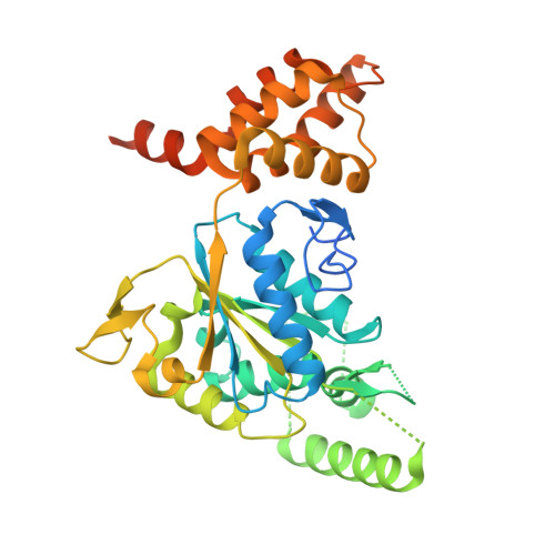
    3UK6

  • PubMed Abstract: 

    The TIP49a and TIP49b proteins belong to the family of AAA+ ATPases and play essential roles in vital processes such as transcription, DNA repair, snoRNP biogenesis, and chromatin remodeling. We report the crystal structure of a TIP49b hexamer and the comparative analysis of large-scale conformational flexibility of TIP49a, TIP49b, and TIP49a/TIP49b complexes using molecular modeling and molecular dynamics simulations in a water environment. Our results establish key principles of domain mobility that affect protein conformation and biochemical properties, including a mechanistic basis for the downregulation of ATPase activity upon protein hexamerization. These approaches, applied to the lik-TIP49b mutant reported to possess enhanced DNA-independent ATPase activity, help explain how a three-amino acid insertion remotely affects the structure and conformational dynamics of the ATP binding and hydrolysis pocket while uncoupling ATP hydrolysis from DNA binding. This might be similar to the effects of conformations adopted by TIP49 heterohexamers.


  • Organizational Affiliation

    Department of Biophysics, Saint Petersburg State Polytechnical University, Saint Petersburg 197376, Russia.


Macromolecules
Find similar proteins by:  (by identity cutoff)  |  3D Structure
Entity ID: 1
MoleculeChains Sequence LengthOrganismDetailsImage
RuvB-like 2
A, B, C, D, E
A, B, C, D, E, F, G, H, I, J, K, L
368Homo sapiensMutation(s): 0 
Gene Names: CGI-46INO80JRUVBL2TIP48TIP49B
EC: 3.6.4.12
UniProt & NIH Common Fund Data Resources
Find proteins for Q9Y230 (Homo sapiens)
Explore Q9Y230 
Go to UniProtKB:  Q9Y230
PHAROS:  Q9Y230
GTEx:  ENSG00000183207 
Entity Groups  
Sequence Clusters30% Identity50% Identity70% Identity90% Identity95% Identity100% Identity
UniProt GroupQ9Y230
Sequence Annotations
Expand
  • Reference Sequence
Small Molecules
Ligands 1 Unique
IDChains Name / Formula / InChI Key2D Diagram3D Interactions
ADP
Query on ADP

Download Ideal Coordinates CCD File 
M [auth A]
N [auth B]
O [auth C]
P [auth D]
Q [auth E]
M [auth A],
N [auth B],
O [auth C],
P [auth D],
Q [auth E],
R [auth F],
S [auth G],
T [auth H],
U [auth I],
V [auth J],
W [auth K],
X [auth L]
ADENOSINE-5'-DIPHOSPHATE
C10 H15 N5 O10 P2
XTWYTFMLZFPYCI-KQYNXXCUSA-N
Experimental Data & Validation

Experimental Data

  • Method: X-RAY DIFFRACTION
  • Resolution: 2.95 Å
  • R-Value Free: 0.264 
  • R-Value Work: 0.241 
  • R-Value Observed: 0.242 
  • Space Group: P 1 21 1
Unit Cell:
Length ( Å )Angle ( ˚ )
a = 113.07α = 90
b = 186.28β = 108.95
c = 129.86γ = 90
Software Package:
Software NamePurpose
DNAdata collection
PHASERphasing
BUSTERrefinement
XDSdata reduction
XSCALEdata scaling

Structure Validation

View Full Validation Report


Ligand Structure Quality Assessment 


Entry History 

Revision History  (Full details and data files)

  • Version 1.0: 2012-07-25
    Type: Initial release
  • Version 1.1: 2012-08-29
    Changes: Database references
  • Version 1.2: 2017-08-23
    Changes: Refinement description, Source and taxonomy
  • Version 1.3: 2024-02-28
    Changes: Data collection, Database references, Derived calculations