3UH2

Tankyrase-1 in complexed with PJ34


Experimental Data Snapshot

  • Method: X-RAY DIFFRACTION
  • Resolution: 2.00 Å
  • R-Value Free: 0.217 
  • R-Value Work: 0.175 
  • R-Value Observed: 0.177 

wwPDB Validation   3D Report Full Report


Ligand Structure Quality Assessment 


This is version 1.1 of the entry. See complete history


Literature

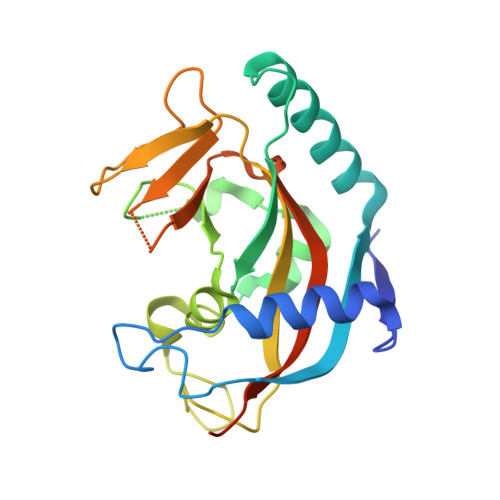
Structure of human tankyrase 1 in complex with small-molecule inhibitors PJ34 and XAV939.

Kirby, C.A.Cheung, A.Fazal, A.Shultz, M.D.Stams, T.

(2012) Acta Crystallogr Sect F Struct Biol Cryst Commun 68: 115-118

  • DOI: https://doi.org/10.1107/S1744309111051219
  • Primary Citation of Related Structures:  
    3UH2, 3UH4

  • PubMed Abstract: 

    The crystal structures of tankyrase 1 (TNKS1) in complex with two small-molecule inhibitors, PJ34 and XAV939, both at 2.0 Å resolution, are reported. The structure of TNKS1 in complex with PJ34 reveals two molecules of PJ34 bound in the NAD(+) donor pocket. One molecule is in the nicotinamide portion of the pocket, as previously observed in other PARP structures, while the second molecule is bound in the adenosine portion of the pocket. Additionally, unlike the unliganded crystallization system, the TNKS1-PJ34 crystallization system has the NAD(+) donor site accessible to bulk solvent in the crystal, which allows displacement soaking. The TNKS1-PJ34 crystallization system was used to determine the structure of TNKS1 in complex with XAV939. These structures provide a basis for the start of a structure-based drug-design campaign for TNKS1.


  • Organizational Affiliation

    Novartis Institute for Biomedical Research, Cambridge, MA 02139, USA.


Macromolecules
Find similar proteins by:  (by identity cutoff)  |  3D Structure
Entity ID: 1
MoleculeChains Sequence LengthOrganismDetailsImage
Tankyrase-1
A, B
224Homo sapiensMutation(s): 1 
EC: 2.4.2.30
UniProt & NIH Common Fund Data Resources
Find proteins for O95271 (Homo sapiens)
Explore O95271 
Go to UniProtKB:  O95271
PHAROS:  O95271
GTEx:  ENSG00000173273 
Entity Groups  
Sequence Clusters30% Identity50% Identity70% Identity90% Identity95% Identity100% Identity
UniProt GroupO95271
Sequence Annotations
Expand
  • Reference Sequence
Small Molecules
Ligands 3 Unique
IDChains Name / Formula / InChI Key2D Diagram3D Interactions
P34
Query on P34

Download Ideal Coordinates CCD File 
D [auth A],
E [auth A],
K [auth B],
L [auth B]
N~2~,N~2~-DIMETHYL-N~1~-(6-OXO-5,6-DIHYDROPHENANTHRIDIN-2-YL)GLYCINAMIDE
C17 H17 N3 O2
UYJZZVDLGDDTCL-UHFFFAOYSA-N
SO4
Query on SO4

Download Ideal Coordinates CCD File 
F [auth A],
G [auth A],
H [auth A],
I [auth A],
M [auth B]
SULFATE ION
O4 S
QAOWNCQODCNURD-UHFFFAOYSA-L
ZN
Query on ZN

Download Ideal Coordinates CCD File 
C [auth A],
J [auth B]
ZINC ION
Zn
PTFCDOFLOPIGGS-UHFFFAOYSA-N
Binding Affinity Annotations 
IDSourceBinding Affinity
P34 BindingDB:  3UH2 IC50: min: 39, max: 2951 (nM) from 6 assay(s)
PDBBind:  3UH2 IC50: 1000 (nM) from 1 assay(s)
Binding MOAD:  3UH2 IC50: 1000 (nM) from 1 assay(s)
Experimental Data & Validation

Experimental Data

  • Method: X-RAY DIFFRACTION
  • Resolution: 2.00 Å
  • R-Value Free: 0.217 
  • R-Value Work: 0.175 
  • R-Value Observed: 0.177 
  • Space Group: C 1 2 1
Unit Cell:
Length ( Å )Angle ( ˚ )
a = 125.26α = 90
b = 43.3β = 91.36
c = 88.54γ = 90
Software Package:
Software NamePurpose
ADSCdata collection
PHASERphasing
BUSTERrefinement
autoPROCdata scaling
SCALAdata scaling

Structure Validation

View Full Validation Report


Ligand Structure Quality Assessment 


Entry History 

Deposition Data

Revision History  (Full details and data files)

  • Version 1.0: 2012-02-15
    Type: Initial release
  • Version 1.1: 2023-09-13
    Changes: Data collection, Database references, Derived calculations, Refinement description