3UG6

Crystal Structure of Get3 from Methanocaldococcus jannaschii


Experimental Data Snapshot

  • Method: X-RAY DIFFRACTION
  • Resolution: 3.30 Å
  • R-Value Free: 0.287 
  • R-Value Work: 0.249 
  • R-Value Observed: 0.251 

wwPDB Validation   3D Report Full Report


Ligand Structure Quality Assessment 


This is version 1.3 of the entry. See complete history


Literature

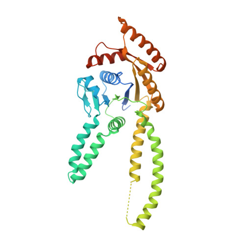
Tail-anchor targeting by a Get3 tetramer: the structure of an archaeal homologue.

Suloway, C.J.Rome, M.E.Clemons, W.M.

(2012) EMBO J 31: 707-719

  • DOI: https://doi.org/10.1038/emboj.2011.433
  • Primary Citation of Related Structures:  
    3UG6, 3UG7

  • PubMed Abstract: 

    Efficient delivery of membrane proteins is a critical cellular process. The recently elucidated GET (Guided Entry of TA proteins) pathway is responsible for the targeted delivery of tail-anchored (TA) membrane proteins to the endoplasmic reticulum. The central player is the ATPase Get3, which in its free form exists as a dimer. Biochemical evidence suggests a role for a tetramer of Get3. Here, we present the first crystal structure of an archaeal Get3 homologue that exists as a tetramer and is capable of TA protein binding. The tetramer generates a hydrophobic chamber that we propose binds the TA protein. We use small-angle X-ray scattering to provide the first structural information of a fungal Get3/TA protein complex showing that the overall molecular envelope is consistent with the archaeal tetramer structure. Moreover, we show that this fungal tetramer complex is capable of TA insertion. This allows us to suggest a model where a tetramer of Get3 sequesters a TA protein during targeting to the membrane.


  • Organizational Affiliation

    Division of Chemistry and Chemical Engineering, California Institute of Technology, Pasadena, CA 91125, USA.


Macromolecules
Find similar proteins by:  (by identity cutoff)  |  3D Structure
Entity ID: 1
MoleculeChains Sequence LengthOrganismDetailsImage
arsenical pump-driving ATPase
A, B, C, D
349Methanocaldococcus jannaschii DSM 2661Mutation(s): 0 
Gene Names: MJ1142
EC: 3.6.3.16
UniProt
Find proteins for Q58542 (Methanocaldococcus jannaschii (strain ATCC 43067 / DSM 2661 / JAL-1 / JCM 10045 / NBRC 100440))
Explore Q58542 
Go to UniProtKB:  Q58542
Entity Groups  
Sequence Clusters30% Identity50% Identity70% Identity90% Identity95% Identity100% Identity
UniProt GroupQ58542
Sequence Annotations
Expand
  • Reference Sequence
Small Molecules
Ligands 3 Unique
IDChains Name / Formula / InChI Key2D Diagram3D Interactions
ADP
Query on ADP

Download Ideal Coordinates CCD File 
E [auth A],
H [auth B],
J [auth C],
M [auth D]
ADENOSINE-5'-DIPHOSPHATE
C10 H15 N5 O10 P2
XTWYTFMLZFPYCI-KQYNXXCUSA-N
ZN
Query on ZN

Download Ideal Coordinates CCD File 
G [auth A],
L [auth C]
ZINC ION
Zn
PTFCDOFLOPIGGS-UHFFFAOYSA-N
MG
Query on MG

Download Ideal Coordinates CCD File 
F [auth A],
I [auth B],
K [auth C],
N [auth D]
MAGNESIUM ION
Mg
JLVVSXFLKOJNIY-UHFFFAOYSA-N
Experimental Data & Validation

Experimental Data

  • Method: X-RAY DIFFRACTION
  • Resolution: 3.30 Å
  • R-Value Free: 0.287 
  • R-Value Work: 0.249 
  • R-Value Observed: 0.251 
  • Space Group: P 1 21 1
Unit Cell:
Length ( Å )Angle ( ˚ )
a = 85.736α = 90
b = 76.874β = 108.828
c = 127.81γ = 90
Software Package:
Software NamePurpose
PHENIXrefinement
Blu-Icedata collection
MOSFLMdata reduction
SCALAdata scaling
PHASERphasing

Structure Validation

View Full Validation Report


Ligand Structure Quality Assessment 


Entry History 

Deposition Data

Revision History  (Full details and data files)

  • Version 1.0: 2011-12-07
    Type: Initial release
  • Version 1.1: 2012-01-11
    Changes: Database references
  • Version 1.2: 2012-02-15
    Changes: Database references
  • Version 1.3: 2024-02-28
    Changes: Data collection, Database references, Derived calculations