3TOJ

Structure of the SPRY domain of human Ash2L


Experimental Data Snapshot

  • Method: X-RAY DIFFRACTION
  • Resolution: 2.07 Å
  • R-Value Free: 0.206 
  • R-Value Work: 0.176 
  • R-Value Observed: 0.179 

wwPDB Validation   3D Report Full Report


This is version 1.2 of the entry. See complete history


Literature

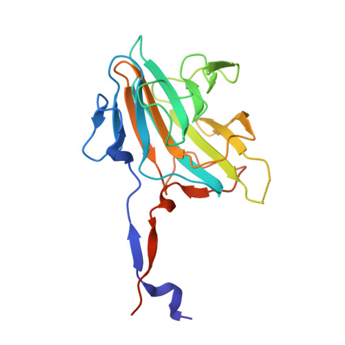
Structure of the SPRY domain of human Ash2L and its interactions with RbBP5 and DPY30.

Chen, Y.Cao, F.Wan, B.Dou, Y.Lei, M.

(2012) Cell Res 22: 598-602


Macromolecules
Find similar proteins by:  (by identity cutoff)  |  3D Structure
Entity ID: 1
MoleculeChains Sequence LengthOrganismDetailsImage
Set1/Ash2 histone methyltransferase complex subunit ASH2
A, B
213Homo sapiensMutation(s): 0 
Gene Names: ASH2LASH2L1
UniProt & NIH Common Fund Data Resources
Find proteins for Q9UBL3 (Homo sapiens)
Explore Q9UBL3 
Go to UniProtKB:  Q9UBL3
PHAROS:  Q9UBL3
GTEx:  ENSG00000129691 
Entity Groups  
Sequence Clusters30% Identity50% Identity70% Identity90% Identity95% Identity100% Identity
UniProt GroupQ9UBL3
Sequence Annotations
Expand
  • Reference Sequence
Experimental Data & Validation

Experimental Data

  • Method: X-RAY DIFFRACTION
  • Resolution: 2.07 Å
  • R-Value Free: 0.206 
  • R-Value Work: 0.176 
  • R-Value Observed: 0.179 
  • Space Group: P 31 2 1
Unit Cell:
Length ( Å )Angle ( ˚ )
a = 115.289α = 90
b = 115.289β = 90
c = 105.78γ = 120
Software Package:
Software NamePurpose
HKL-2000data collection
SHARPphasing
PHENIXrefinement
HKL-2000data reduction
HKL-2000data scaling

Structure Validation

View Full Validation Report


Entry History 

Deposition Data

Revision History  (Full details and data files)

  • Version 1.0: 2012-01-25
    Type: Initial release
  • Version 1.1: 2012-07-18
    Changes: Database references
  • Version 1.2: 2017-08-23
    Changes: Refinement description, Source and taxonomy