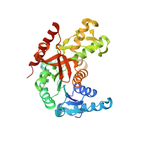
Crystal structure of Bacillus anthracis str. Ames malate dehydrogenase in closed conformation.
Blus, B.J., Chruszcz, M., Tkaczuk, K.L., Osinski, T., Cymborowski, M., Kudritska, M., Grimshaw, S., Savchenko, A., Anderson, W.F., Minor, W.To be published.